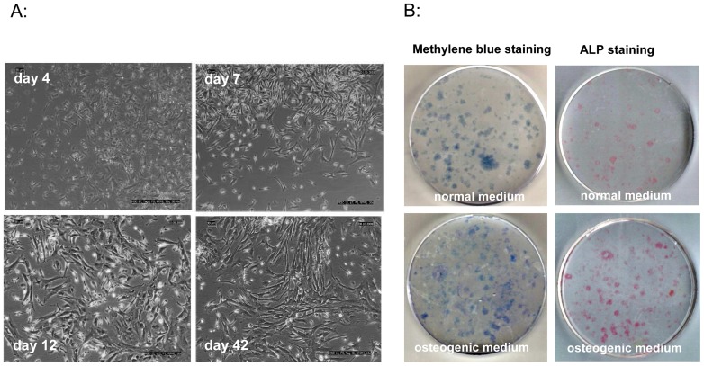
Figure 5. MSC characterization.
A: MSCs were evaluated under a light microscope and representative photographs were captured on days 4, 7, 12, and 42 of incubation. Cells show typical fibroblast morphology and undergo typical time-dependent transitions among three morphologically distinct cell types: thin spindle-shaped cells, wider spindle-shaped cells, and still wider spindle-shaped cells. B: Fibroblastic colony-forming unit assays (CFU-F) were performed for 14 days. Colonies were then stained for ALP and methylene blue. When cultivated in normal medium, only a few colonies expressed ALP (line 1), whereas nearly all of the colonies stained with methylene blue also showed ALP expression when cultivated in osteogenic medium (line 2).

