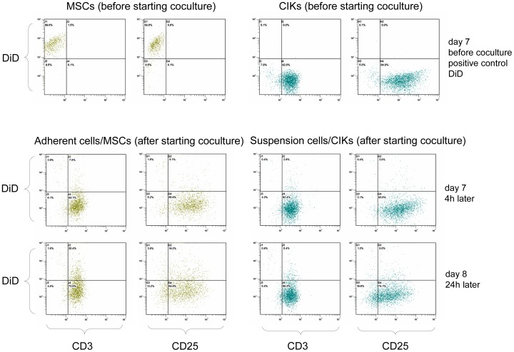
Figure 8. DiD-labelled MSCs.
To investigate whether cocultured MSCs were differentiated, we carried out coculture experiments using DiD-labelled MSCs. Suspended cells (cCIKs) and adherent cells were harvested separately from each other after 4 and 24 h of coculture, stained with anti-CD3 and anti-CD25, and analysed using flow cytometry. The first line shows the results of flow cytometry analysis just before the addition of CIKs to the labelled MSCs. Clearly positive DiD signals were seen at the MSCs (positive control). However, CD3 and CD25 were negative, which was expected for MSCs. The CIKs were not labelled and were therefore DiD negative. As for CIKs characteristic, CD3 and CD25 were positive. The second line shows the results of FACS analysis of adherent cells and suspended cells after 4 h of coculture. Adherent cells have switched phenotype from CD3−/CD25− to CD3+/CD25+. DiD signals of adherent cells have disappeared. Suspended cells were still CD3+/CD25+ and DiD−. The third line (after 24 h) confirms the results seen after 4 h. Adherent cells showed the phenotype of the suspended cells and were DiD−.

