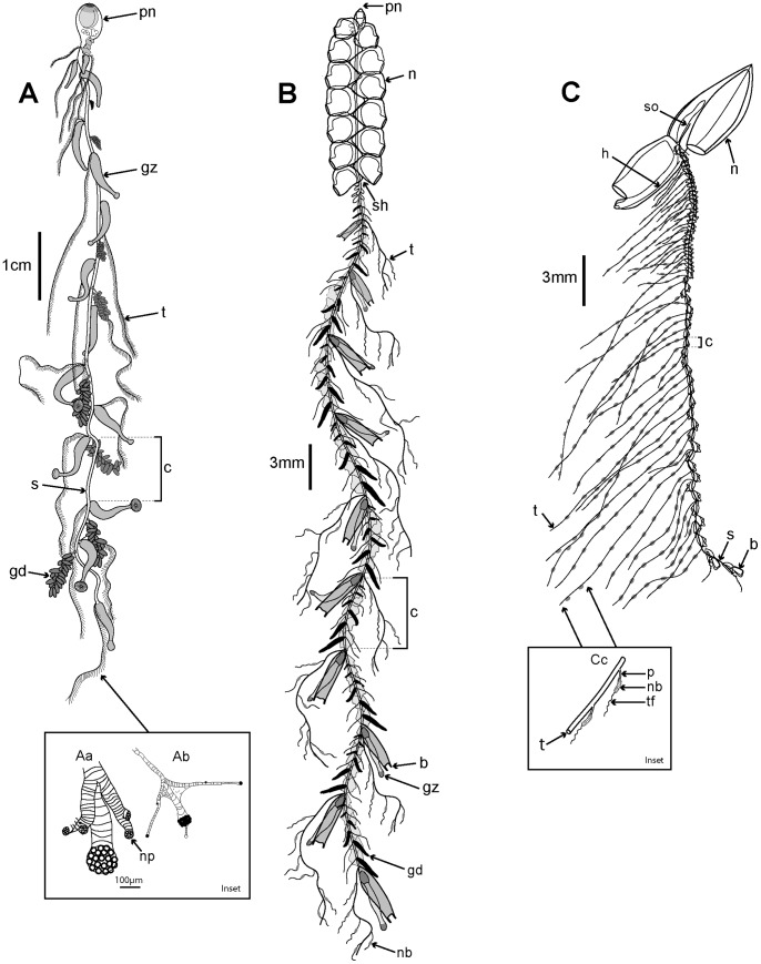
Figure 3. Three typical siphonophore body plans.
A. Long-stemmed cystonect Rhizophysa eysenhardti (derived from [66] pl. 14 fig. 1): inset shows nematocyst pads on two interpretations of tricornuate tentacular side branches from Rhizophysa filiformis, (Aa: derived from [67] fig. 5 and Ab: derived from [9] pl. 4, fig. 2): B. Long-stemmed physonect Nanomia bijuga (derived from [68], pl. 7, fig. 1); C. Typical calycophoran Lensia conoidea (derived from photo image by Rob Sherlock - shown in Fig. 5C): inset Cc shows two tentilla attached to one tentacle (derived from [69] pl. 11, fig. 2). Labels: b - bract; c – cormidium; gd - gonodendron; gz - gastrozooid; h – hydroecium; n – nectophore (swimming bell); nb – nematocyst battery (a coiled cnidoband); np – nematocyst pad; p - pedicel; pn – pneumatophore (float); s – stem; sh – siphosomal horn; so – somatocyst; t – tentacle; tf – terminal filament.

