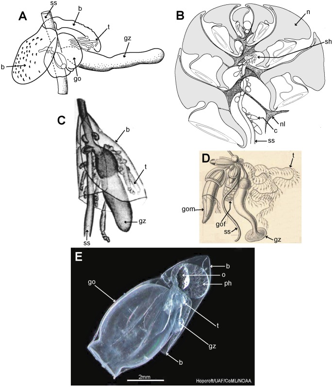
Figure 8. Calycophoran cormidia.
A: Rosacea cymbiformis cormidium (after [6] fig. 2D); B. Hippopodius hippopus section through colony (adapted from [31] fig. 11, [78] txt fig. 13 and [27] fig. 44b); C: Chelophyes appendiculata cormidium (from [34] pl. 11, fig. 1); D. Hippopodius hippopus cormidium; note, no bracts (from [26] pl. 29, fig. 1 in part); E. Dimophyes arctica eudoxid (Russ Hopcroft, UAF). Labels: b – bract, c – cormidium; go – gonophore; gof – female gonophore; gom – male gonophore; gz – gastrozooid; n – nectophore; nl – nectophoral lamella; o – oil globule (in phyllocyst); ph – phyllocyst; sh – siphosomal horn; ss – siphosomal stem; t – tentacle with tentilla.

