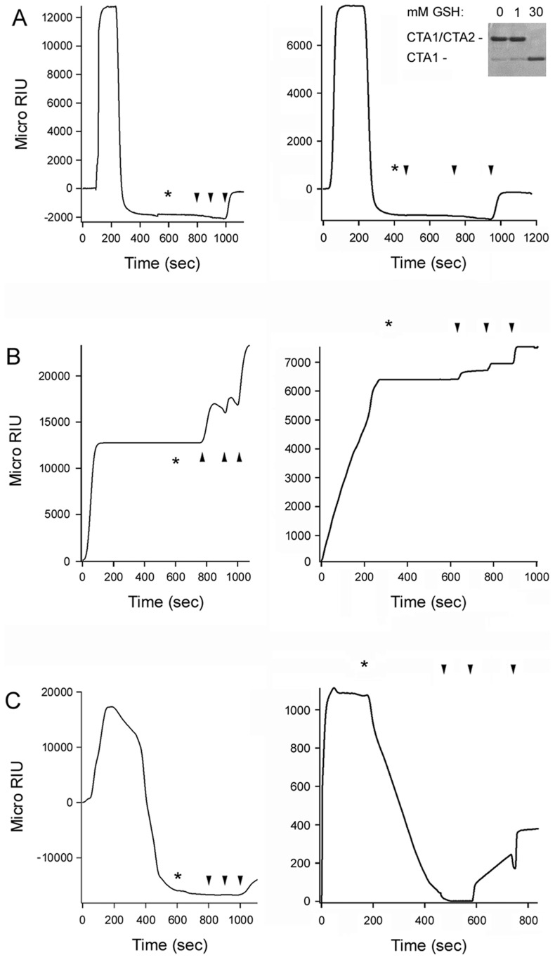
Figure 5. PDI unfolding but not oxidoreductase activity is required for disassembly of the CT holotoxin.
SPR was used to monitor the real-time PDI-mediated disassembly of CT. A baseline measurement corresponding to the mass of the sensor-bound CT holotoxin established the 0 MicroRIU signal. The time course was then initiated with perfusion of PDI (A), EDC-treated PDI (B), or bacitracin-treated PDI (C) over the CT-coated sensor. The perfusion buffer contained either 30 mM GSH (left panels) or 1 mM GSH (right panels); non-reducing SDS-PAGE with Coomassie staining found the CTA1/CTA2 disulfide bond was reduced at 30 mM GSH but not 1 mM GSH (inset, right panel of A). PDI was removed from the perfusion buffer at time intervals denoted by asterisks and was replaced with sequential additions of anti-PDI, anti-CTA1, and anti-CTB antibodies as indicated by the arrowheads. One of two representative experiments is shown for each condition.

