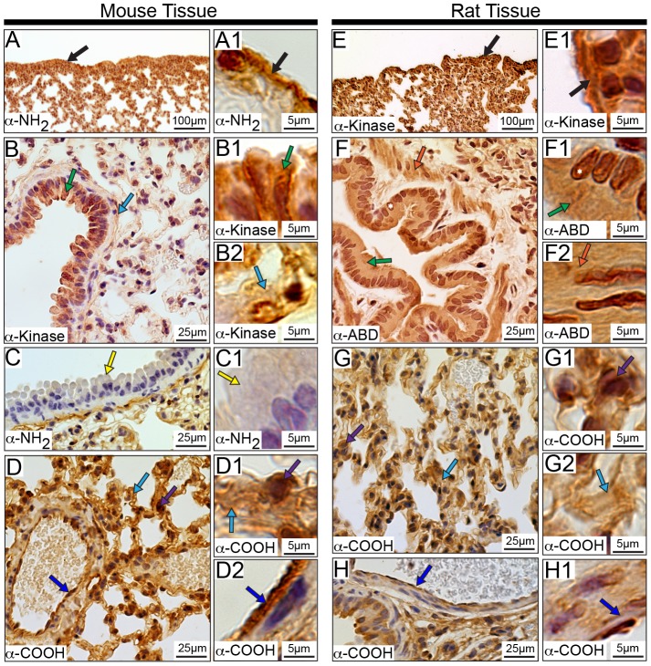
Figure 14. Distribution of obscurins in rodent lung.
Obscurins localize to the mesothelial cells making up the pleura, which surrounds the lung (A-A1 and E-E1, mouse and rat, respectively; black arrows). Within the bronchioles, particular obscurins are found in the cytoplasm of Clara cells (green arrows) of both mouse (B-B1) and rat (F-F1) lung. Interestingly, obscurins carrying the NH2-terminal epitopes are absent from the cytoplasm of Clara cells in mouse tissue (C-C1, yellow arrows). Obscurins are also found in the fibrous component of the connective tissue (B, B2, D-D1, G, and G2, light blue arrows) as well as in cells within the connective tissue (purple arrow) in both mouse (D-D1) and rat (G-G1) lung. Similar to observations in other tissues, obscurins localize to VECs (dark blue arrows) in the mouse (D and D2) and rat (H-H1) lung. Smooth muscle within surrounding the bronchioles contains obscurins only in rat tissue (F and F2, orange arrows). Images are shown at multiple magnifications to highlight the various immunopositive structures. Scale bars are included in each panel for reference.

