FIGURE 2.
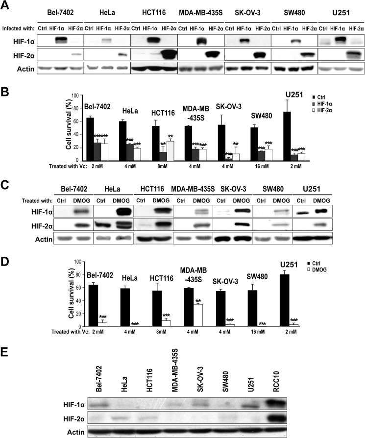
Activation of HIF renders non-renal VHL-competent cancer cells more sensitive to Vc-induced toxicity. A, representative Western blot analysis for HIFα subunits using lysates from non-renal VHL-competent cancer cell lines overexpressing constitutively active forms of HIF1α and HIF2α showing the expected increase in protein levels. Actin was used as a loading control (Ctrl). Lentiviruses producing GFP were used as a control. B, overexpression of HIF1α and HIF2α increases Vc-induced toxicity in non-renal VHL-competent cancer cell lines. Vc was added for 1 h to all cell lines (also in D). The concentrations were 2 mm for Bel-7402 and U251, 4 mm for HeLa, MDA-MB-435S and SK-OV-3, 8 mm for HCT116, and 16 mm for SW480 cells. Cell survival (percent) was measured by counting viable cells and is represented as relative to untreated cells (also in D). The mean ± S.D. of four independent experiments is shown (also in D). C, representative Western blot analysis for HIFα subunits using lysates from non-renal VHL-competent cancer cell lines treated or not treated with 0.5 mm DMOG for 24 h. D, pretreatment with 0.5 mm DMOG for 24 h increases Vc-induced toxicity in non-renal VHL-competent cancer cell lines. E, representative Western blot analysis comparing the basal levels (in normoxia) of HIF1α and HIF2α in a panel of non-renal VHL-competent cancer cell lines. Lysates from VHL-defective RCC10 cells were used as a control. **, p < 0.01; ***, p < 0.001.

