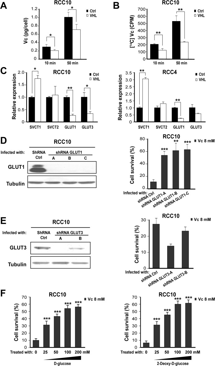
FIGURE 3.
Increased DHA transport through GLUT1 underlies Vc-induced toxicity in VHL-defective renal cancer cells. A, HPLC shows increased intracellular Vc levels in VHL-defective RCC10 cells overexpressing VHL or the empty vector. Vc was added for 10 min or 50 min (also in B). The mean ± S.D. of four independent experiments is shown. Ctrl, control. B, liquid scintillation values after adding isotope-labeled Vc to VHL-defective RCC10 cells overexpressing VHL or the empty vector. The mean ± S.D. of three independent experiments is shown. C, representative quantitative PCR analysis showing the relative expression of different Vc transporters in VHL-defective RCC10 and RCC4 overexpressing VHL or the empty vector. D, left panel, representative Western blot analysis showing reduction of GLUT1 protein after transduction of VHL-defective RCC10 cells with three shRNA constructs (A, B, and C). Right panel, shRNA for GLUT1 reduces Vc-induced toxicity in VHL-defective RCC10 cells. Vc was added for 1 h (also in E and F). Cell survival (percent) was measured by counting viable cells and is represented as relative to untreated cells (also in E right and F). The mean ± S.D. of three independent experiments is shown. E, similar experiments as in D using two shRNA vectors (A and B) for GLUT3, which fail to reduce Vc-induced toxicity in VHL-defective RCC10 cells. The mean ± S.D. of four independent experiments is shown (also in F). F, 2-deoxy-d-glucose and d-glucose reduce Vc-induced toxicity in VHL-defective RCC10 cells. The two compounds were added simultaneously with Vc. *, p < 0.05; **, p < 0.01; ***, p < 0.001.

