FIGURE 2.
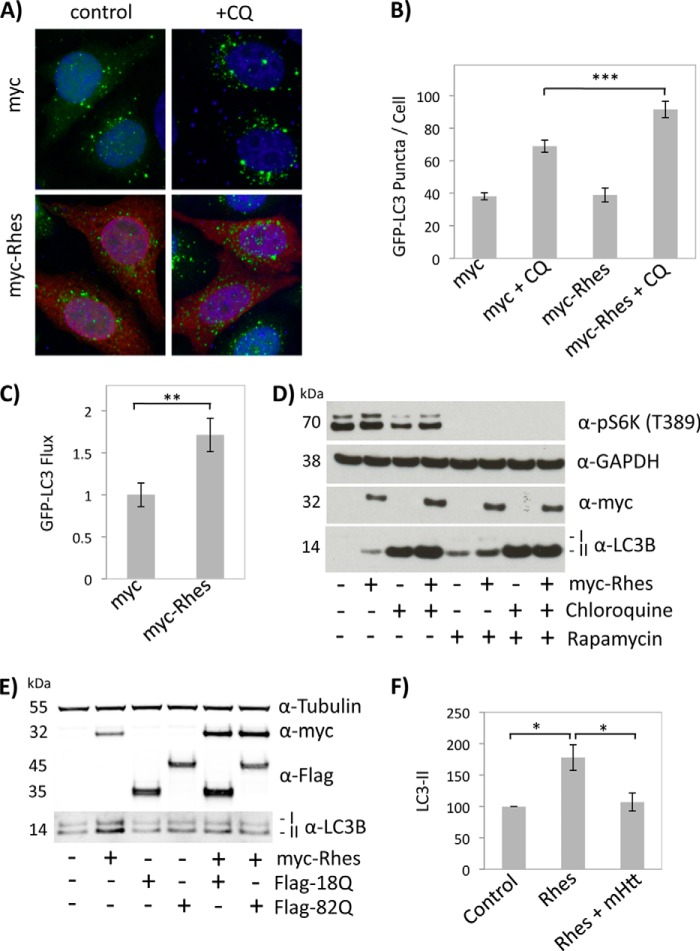
Rhes activates autophagy in HeLa cells independent of mTOR. A, HeLa cells stably expressing GFP-LC3 were transfected with Myc or Myc-Rhes for 24 h, transferred to low autophagy medium for 24 h, and then treated with or without chloroquine (CQ) for 2 h. Representative confocal images show GFP-LC3 (green), anti-Myc immunofluorescence (red), and Hoescht 33324 nuclear stain (blue). B, GPF-LC3 puncta analysis from HeLa cells in A demonstrates that Rhes increases the number of GFP-LC3 puncta following chloroquine treatment, consistent with increased autophagic flux. C, GFP-LC3 flux following 2 h of chloroquine treatment in cells transfected with Myc or Myc-Rhes, calculated as the number of GFP-LC3 puncta in the presence of CQ minus the number of GFP-LC3 puncta without CQ. D, Overexpression of Myc-Rhes in HEK293 cells increases LC3-II levels in the presence of rapamycin. Cells were transfected for 48 h and then placed in medium ± CQ and ± rapamycin for 90 min. E, huntingtin prevents Rhes-induced autophagy. HEK293 cells were transfected with control vectors or Myc-Rhes and wtHtt (FLAG-Htt-N171–18Q) or mHtt (FLAG-Htt-N171–82Q) for 24 h. F, quantification of LC3-II levels in HEK293 cells expressing control vector, Rhes alone, or both Rhes and mHtt from three separate experiments. *, p < 0.05; **, p < 0.01; ***, p < 0.001.

