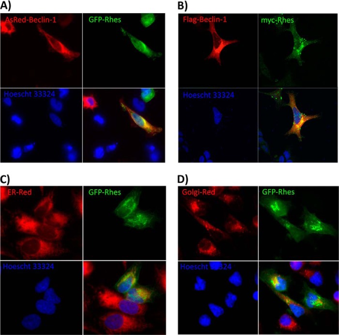
FIGURE 4.
Rhes co-localizes with Beclin-1. A, cDNA for AsRed-Beclin-1 and GFP-Rhes were transfected into HEK293 cells and expressed for 24 h and then fixed with 4% paraformaldehyde and imaged using fluorescence microscopy. B, FLAG-Beclin-1 and Myc-Rhes were transfected into HEK293 cells and expressed for 24 h and then fixed with 4% paraformaldehyde and imaged following immunofluorescence labeling with primary antibodies for the indicated epitope tag and fluorescently labeled secondary antibodies. ER-Red (glibenclamide BODIPY-TR) (C) and Golgi-Red (BODIPY-TR ceramide) (D) were added to HeLa cells transfected with GFP-Rhes as recommended by Invitrogen, and live cells were imaged in PBS. Expression in the endoplasmic reticulum (ER) and trans-Golgi network (TGN) is consistent with the previously reported localization of Beclin-1. Nuclei are stained with Hoescht 33324.

