Abstract
Background
Delay of as much as 5 months between ACL injury and surgery is known to be associated with increased risk of a medial meniscal tear, but the risk of additional meniscal tear progression with a longer delay to surgery is unclear.
Questions/purposes
We determined the (1) times of injury, MRI, and surgery in adolescents with ACL tears, and whether (2) timing of surgery, or (3) initial integrity of the meniscus seen on MR images predicted development of meniscal tears.
Methods
We reviewed 112 adolescents who were 15 ± 1 years old (mean ± SD) (range, 11–16 years) with a torn ACL. These patients underwent surgical repair from 2005 to 2011 in a Canadian city. We compared dates of injury, MRI, and surgery. A pediatric and musculoskeletal fellowship-trained radiologist reread the MR images, and meniscal injuries were graded according to severity. This was compared with surgical findings described in the operative report.
Results
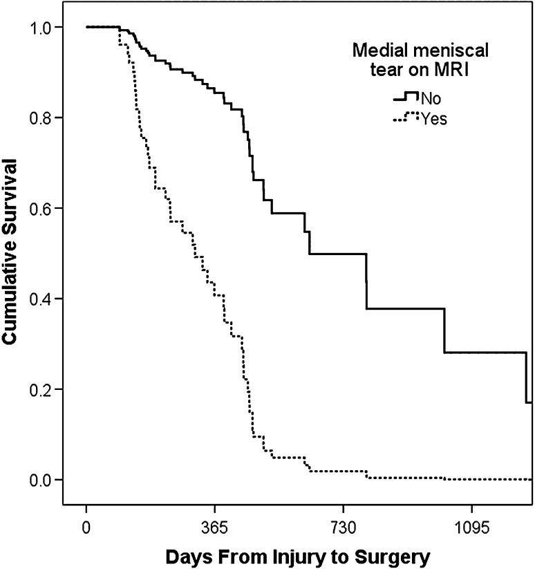
Time after injury to MRI and surgery averaged 77 days (range, 1–377 days) and 342 days (range, 42–1637 days), respectively. Patients with new or worsened medial meniscal tears had waited longer for surgery (445 versus 290 days; p = 0.002). Bucket handle medial meniscal tears were more common in patients with surgery more than 1 year after injury than others (15 of 34 versus 14 of 75; p = 0.013). A medial meniscal tear observed on MR images was a significant covariate for a torn meniscus at surgery (relative risk, 5.7; 95% CI, 2.8–11.6). Medial meniscal survival continued to decline sharply greater than 1 year after injury.
Conclusions
Medial meniscal tears, especially bucket handle tears, increased steadily in frequency more than 1 year after ACL injury. Timely ACL reconstruction may be warranted to reduce the risk of further medial meniscal damage even in patients whose original injury occurred more than 1 year before.
Level of Evidence
Level IV, prognostic study. See the Instructions for Authors for a complete description of levels of evidence.
Introduction
Tears of the ACL are becoming more common in the adolescent population [5, 38], with the annual incidence estimated at 16 per 1000 high school students [3]. This represents a high burden of future disability, given that ACL tears in childhood are associated with a high risk of early osteoarthritis (OA). Radiographic OA is 105 times more likely to occur in adults who had childhood ACL tears [36]. Abnormal joint forces in an ACL-deficient knee are associated with increased risk of injury to menisci, which normally transmit load and absorb shocks between the femur and tibia [37]. With each episode of giving way, meniscal tears can become more complex and less amenable to repair [8]. Studies in adult patients [7, 8, 24, 39] and in pediatric patients [12, 28, 32] have shown an increased incidence of medial meniscal tears with delayed ACL reconstruction. Although the treatment of ACL tears in skeletally immature patients remains controversial [33], the risk of physeal injury and resulting growth disturbance from ACL repair to a patient with open physes is now thought to be small [15, 22], and many centers (including our institution) opt for prompt reconstruction before skeletal maturity [9, 16, 20, 25, 31, 34, 40].
Previous American studies showing an increased incidence of meniscal tears with delay to ACL reconstruction in adolescent patients have looked at relatively short-term tear progression (< 5 months) [12, 28, 32], but the longer-term prognosis is unclear. This is important in cases of initially missed ACL tears in patients who present late and in the Canadian public healthcare system where delays for imaging and surgery are substantially longer than in the United States [14].
In this study, we therefore compared longer-term patterns of evolution of meniscal tears between time of MRI and surgery in pediatric patients with ACL tears. We specifically determined (1) the time from injury to MRI to surgery in Canadian adolescents with ACL tears and whether (2) the timing of surgery or (3) the initial integrity of the meniscus observed on MR images predicted the development of meniscal tears, particularly bucket handle tears.
Patients and Methods
Ethical approval for this study was obtained from our institutional health research ethics board. The requirement for consent was waived for this retrospective study. We performed a retrospective search of MRI knee studies performed on 10- to 16-year-old patients from July 2005 to September 2011 at two local hospitals and a private MRI center. This identified 1160 patients. We then performed a focused chart review to identify the patients who went on to have ACL reconstruction (120 patients). Exclusion criteria included poor-quality scans (three patients) and operative reports not available for review (eight patients). This identified 112 patients with a mean age of 15.4 years (SD, 1.0) at the time of injury (Table 1).
Table 1.
Demographics of the patients
| Group | Number of patients | Age (years)* | Sex (male/female) (number of patients) |
|---|---|---|---|
| Total | 112 | 15.4 ± 1.0 (11.5–16.9) | 51 (46%) / 61 (54%) |
| Open physes† | 48 (43%) | 14.7 ± 1.0 (11.5–16.4) | 29 (60%) / 19 (40%) |
| Closed physes | 64 (57%) | 15.9 ± 0.8 (13.8–16.9) | 22 (34%) / 42 (66%) |
* Values are expressed as mean ± SD (range); †there was a statistically significant difference in the occurrence of open physes between boys (56.9%) and girls (31.1%) (p = 0.01).
During the period in question, at the three participating centers, we generally recommended that adolescents with ACL tears receive MRI within 4 to 6 weeks of injury. In discussion with treating orthopaedic surgeons, the preference was to have ACL reconstruction on an adolescent patient performed within 12 weeks of injury. However, given limited availability in the Canadian healthcare system, a more realistic goal for surgery was within 5 months of injury.
The referrals for requested imaging were reviewed along with any other available patient information to extract patient age, sex, date of injury, and date of imaging. A dual-fellowship-trained pediatric/musculoskeletal radiologist (JLJ), who did not have access to the initial imaging report and was blinded to any clinical and surgical findings, reread the MR images to assess the ACL, menisci, and bony structures. Meniscal injuries were graded on a scale from 1 to 4: 1 = trivial tear, very short, peripheral; 2 = substantial tear but undisplaced; 3 = displaced portion or involvement of meniscal root; 4 = bucket handle meniscal tear (Fig. 1). Isolated meniscocapsular junction injuries were noted but graded 0. MRI studies were obtained on 1.5 T Siemens (Erlangen, Germany) and General Electric (Milwaukee, WI, USA) machines. The sequences included sagittal and coronal proton density, sagittal proton density fat-saturated, coronal short tau inversion recovery, and axial proton density fat-saturated or short tau inversion recovery sequences, with a slice thickness of 3 to 4 mm and a field of view of 160 x 160 mm. Information regarding date of surgery, type of surgery performed, and any injuries identified intraoperatively were obtained from patient charts. The surgeries performed were all arthroscopic ACL reconstructions (using either semitendinosus/gracilis grafts or patellar tendon grafts) and performed at five centers by eight orthopaedic surgeons.
Fig. 1.
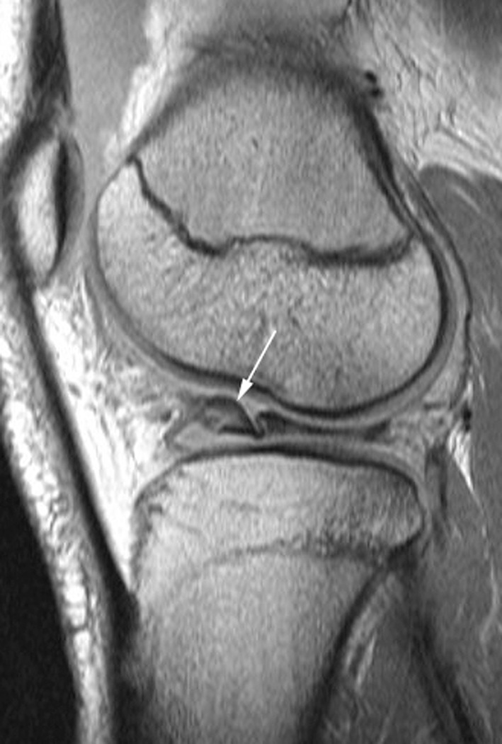
A sagittal fat-saturated proton density MR image show a bucket handle tear (arrow) of the lateral meniscus with a folded displaced position in a 16-year-old patient.
Statistical analysis was performed using Microsoft® Excel® (Version 2010; Redmond, WA, USA) and SPSS® (Version 9; Chicago, IL, USA) software. We tested differences between means of continuous variables using Student’s t-test and difference between proportions of binary variables using Fisher’s exact test. One-way mixed-methods ANOVA was performed to test whether a poor meniscal outcome (tear occurring after initial MRI or bucket handle tear) was associated with longer time to surgery. In an analysis similar to that performed by Dumont et al. [12] (who used a 150-day threshold), we then compared meniscal tears in early versus late reconstructions using a 1-year postinjury threshold. The late-surgery cohort (surgery greater than 1 year after initial injury, n = 37 of 112) was compared with the early-surgery cohort (surgery less than 1 year postinjury, n = 75 of 112) using chi-square tests to assess proportions of binary variables. We also performed survival analysis using Cox proportional-hazard regression modeling at the medial and lateral meniscus to assess meniscal survival to surgery as it related to meniscal integrity at initial MRI and other covariate factors (patient sex, age at injury, whether physes were open or closed, whether effusion or high-grade non-ACL ligamentous injury was present at MRI). Values were considered statistically significant at probability values less than 0.05.
Results
Time from Injury to MRI to Surgery
Canadian adolescents had to wait substantial periods of time to receive diagnostic imaging and definitive surgical treatment (Table 2). Imaging was done an average of 77 days after injury, with 94 patients receiving MRI within 3 months (84%) and seven patients greater than 6 months (6%). On MR images, 108 complete tears of the ACL (Fig. 2) and four high-grade tears were seen. At the time of surgery, all patients were found to have complete ACL tears. The mean time from injury to surgery was 342 days, occurring within 4 months in eight patients (7%), in 4 to 12 months in 67 patients (60%), from 1 to 2 years in 28 patients (25%), and in more than 2 years in nine patients (8%). The mean time from MRI to surgery was 265 days, with 103 patients (92%) undergoing surgery within 1 year of imaging. There was no meaningful correlation between time to MRI and time to surgery (R2 = 0.01). It has been shown that patients with ACL tears typically present to physicians within 24 hours [18], so it is thought that the delays to MRI and surgery were not affected substantially by delays to presentation.
Table 2.
Time from injury to MRI to surgery
| Period | Mean (days) | Standard deviation (days) | Range (days) |
|---|---|---|---|
| Injury to MRI | 77 | 71 | 1–377 |
| Injury to surgery | 342 | 258 | 42–1637 |
| MRI to surgery | 265 | 260 | 16–1625 |
Fig. 2.
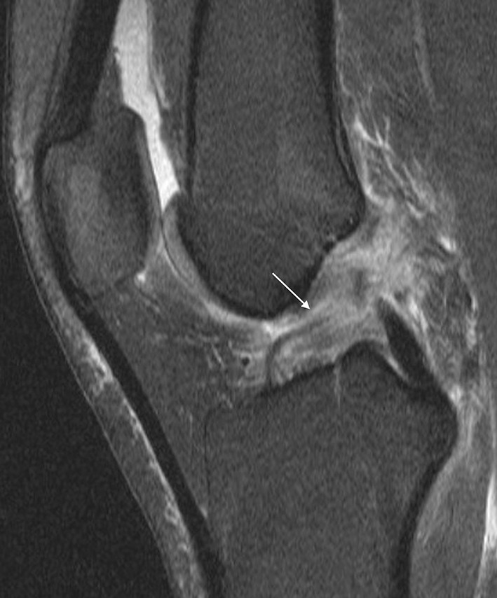
A complete ACL tear (arrow) with a moderate-sized joint effusion in a 16-year-old girl is shown on a sagittal fat-saturated proton density MR image.
Timing of Surgery and Risk of Meniscal Tear
A longer interval between injury and surgery was associated with an increased likelihood of new or worsened medial meniscal tears, particularly medial bucket handle tears. When comparing the medial and lateral menisci at MRI and surgery, new or higher-grade tears were seen intraoperatively in 34 of 112 (30%) medial menisci and 35 of 112 (31%) lateral menisci, respectively. For the medial meniscus, patients with new or higher-grade tears had a longer time to surgery than those whose menisci did not worsen (445 versus 290 days, p = 0.002). This relationship was not found with respect to the lateral meniscus (353 versus 311 days, p = 0.45), or when looking specifically at bucket handle tears in either meniscus (medial, 412 versus 315 days, p = 0.08; lateral, 410 versus 331 days, p = 0.31). When comparing early (less than 1 year after injury) versus late (greater than 1 year after injury) surgery cohorts, new or higher-grade medial meniscal tears were significantly more common in the late-surgery group (19 of 37 [51%] versus 15 of 75 [20%], p = 0.001; odds ratio, 4.22; 95% CI, 1.8–9.9). Similarly, the incidence of medial meniscus bucket handle tears was significantly higher in the late-surgery group than in the early-surgery group (15 of 34 [44%] versus 14 of 75 [19%], p = 0.0013). There was no difference in severity of all lateral meniscal tears (10 of 37 [27%] early versus 25 of 75 [33%], p = 0.93) or lateral bucket handle tears (six of 37 [16%] versus seven of 75 [9%], p = 0.29) when comparing the early and late surgery groups (Table 3).
Table 3.
Overview of meniscal injuries
| Type of meniscal injury | Number of tears | |
|---|---|---|
| At MRI | At surgery | |
| Overall | 84 (75%) | 95 (85%) |
| Medial meniscus | 54 (48%)* | 60 (54%) |
| Lateral meniscus | 53 (47%) | 64 (57%) |
| Bucket handle tear of the medial meniscus | 4 | 29 |
| Bucket handle tear of the lateral meniscus | 1 | 13 |
* Nearly 1/2 of these tears (21 of 54) were vertical peripheral tears of the posterior 1/3 of the meniscus or tears of the adjacent meniscocapsular junction and represented nine of the 12 medial meniscal tears seen on MR images that were not identified or had improved by the time of surgery.
Integrity of Meniscus on MRI as Predictor of Tear Worsening at Surgery
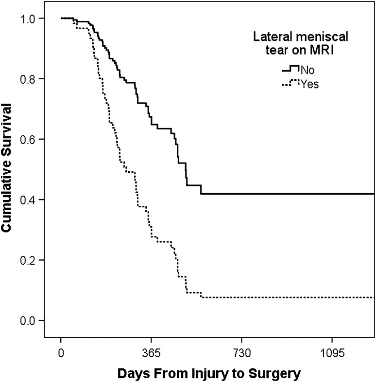
The presence of a meniscal tear observed on the initial MR images was strongly associated with the finding of a meniscal tear at the time of surgery (Table 4). For the medial meniscus, a tear observed on MR images gave a relative risk of 5.7 (95% CI, 2.8–11.6) for a tear observed at arthroscopy. Sex was also a positive predictor, with female sex having a relative risk of 2.1 (95% CI, 1.1–4.2) for a torn meniscus at surgery. Medial meniscal survival continued to decline sharply between 1 and 2 years after injury (Fig. 3). Similarly, the presence of a lateral meniscal tear observed on MR images gave a relative risk of 3.0 (95% CI, 1.7–5.2) of having a torn meniscus at the time of surgery. No other factors (ie, sex) were significant covariates. Lateral meniscal survival declined less steeply and showed less progression than for the medial meniscus in patients who had very late surgery (Fig. 4).
Table 4.
Effect of tear type on progression between MRI and surgery
| Meniscus | Grade of tear* | Incidence on MRI (number of tears) | Surgical findings (number of tears in each category) | Change between MRI and surgery | |||||
|---|---|---|---|---|---|---|---|---|---|
| Improved grade | Same grade | Progressed (higher grade) | New tear | Bucket handle tear at surgery | Improved tear at surgery (%) | Worse tear at surgery (%) | |||
| Medial | No tear | 58 | 0 | 44 | 4 | 10 | 10 | 0 | 24 |
| All tears | 54 | 12 | 22 | 20 | 0 | 19 | 22 | 37 | |
| Grade 1 | 21 | 9 | 5 | 7 | 0 | 4 | 43 | 33 | |
| Grade 2 | 24 | 3 | 11 | 10 | 0 | 9 | 13 | 42 | |
| Grade 3/4 | 9 | 0 | 6 | 3 | 2 | 6 | 0 | 33 | |
| Root tear | 42 | 9 | 22 | 9 | 2 | 6 | 21 | 26 | |
| Lateral | No tear | 59 | 0 | 41 | 0 | 18 | 4 | 0 | 31 |
| All tears | 53 | 9 | 27 | 14 | 3 | 9 | 17 | 32 | |
| Grade 1 | 19 | 2 | 12 | 2 | 3 | 2 | 11 | 26 | |
| Grade 2 | 22 | 4 | 9 | 9 | 0 | 3 | 18 | 41 | |
| Grade 3/4 | 12 | 3 | 6 | 3 | 0 | 4 | 25 | 25 | |
| Root tear | 10 | 4 | 3 | 3 | 0 | 1 | 40 | 30 | |
* Grade 1 = trivial tear (very short, peripheral); 2 = substantial tear but undisplaced; 3 = displaced portion or involvement of meniscal root; 4 = bucket handle meniscal tear.
Fig. 3.
The medial meniscus survival curve is shown. Medial meniscal survival declined sharply during the first year after injury and continued to do so between 1 and 2 years after injury.
Fig. 4.
The lateral meniscus survival curve is shown. Lateral meniscal survival declined less steeply and showed less progression than for the medial meniscus in patients who had very late surgery.
Discussion
Previous American studies of adolescent patients with ACL tears showed an association between delay from initial injury to surgical reconstruction and an increase in medial meniscus tears [12, 28, 32]. However, these studies looked at relatively short intervals (all less than 5 months). In the Canadian healthcare system the delays to imaging and orthopaedic surgery are substantially longer, allowing us to look at meniscal tear progression during greater intervals. The purpose of our study was to identify the wait times to imaging and surgery faced by adolescent patients in Canada after ACL injury and determine if surgical delay beyond 5 months is associated with new or worsened meniscal disorders.
In our series of 112 pediatric patients, the patients had substantial wait times to diagnostic imaging and surgical repair of the torn ACL. Three American studies evaluated similar populations using much shorter times. Millett et al. had a mean time to surgery of 101 days [32], at which point only 4% of our patients had undergone repair. Dumont et al. reported 65% of patients had surgery 150 days or less after injury [12], compared with 20% in our series. Perhaps most striking, Lawrence et al. reported 59% of patients had ACL reconstruction within 12 weeks of injury [28], at which point only 3% of our patients had received surgery. These delays will vary across Canada and even in the same province depending on funding levels, however Alberta has the second highest healthcare expenditure per capita among Canadian provinces, so it is possible that these delays could be worse in other provinces [6]. These findings highlight the significant discrepancies between healthcare delivery in North America and show how our study provides insight into the natural evolution of meniscal injury in the ACL-deficient knee a longer time after injury than otherwise might be possible.
Our study confirmed an increased likelihood of medial meniscal tears when surgery was more than 1 year after injury, with no change in lateral meniscal tears with time. Similar findings have been reported, with Millett et al. [32] using a cut-off of 6 weeks after ACL tear, Lawrence et al. [28] using 12 weeks, and Dumont et al. [12] using 5 months. The increase in medial meniscal tears with time was primarily attributable to the development of new displaced bucket handle meniscal tears. Approximately 1/3 of our patients (37 of 112) had new bucket handle meniscal tears develop between MRI and surgery. As displaced meniscal tears are readily identified on MR images [4, 11, 13], this suggests true progression of medial meniscal tearing related to increased time after injury, probably owing to ongoing instability of the ACL-deficient joint. This is clinically relevant because bucket handle meniscal tears have a relatively poor prognosis, with successful repair in only 59% of pediatric patients with a torn ACL compared with 84% for simple tears [26]. Outcomes were even worse in our study, with only 26.2% of bucket handle meniscal tears successfully repaired based on operative reports. In pediatric patients, meniscal repair is thought to be preferable to partial meniscectomy to reduce the risk of future degenerative changes [35]. An increase in the incidence of bucket handle meniscal tears represents an increase in irreparable meniscal tears that predisposes this subset of patients to OA. Peripheral tears of the posterior 1/3 of the medial meniscus or meniscocapsular junction (Fig. 5) were seen in 21 patients at the time of MRI, accounting for nearly 40% of medial meniscal tears. At the time of surgery, more than 40% of these tears were not seen likely owing to stability of small low-grade tears seen at MRI on surgical probing, and to interval healing as these occur in well-vascularized areas and are known to heal spontaneously [1, 2, 17, 19, 21, 23, 41]. Additionally, the operating surgeon may not have noticed or commented on smaller peripheral tears at the time of arthroscopy. Together, these help to explain why the prevalence of medial meniscal tears observed on MR images in our study (75% at an average of 11 weeks after injury) is substantially greater than the 26% observed by Millett et al. [32] at surgery an average of 14 weeks after injury.
Fig. 5.
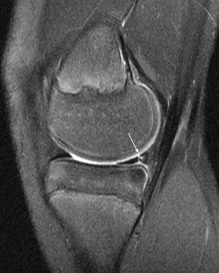
A typical peripheral vertical tear (arrow) at the posterior 1/3 of medial meniscus near the meniscocapsular junction is shown on a sagital proton density fat-saturated MR image in a 15-year-old patient with a complete ACL tear.
Overall, meniscal disorders increased in severity in 1/3 of patients between MRI (mean, 77 days after injury) and surgery (mean, 342 days after injury). Our survival analysis showed that the presence of a meniscal tear observed on MR images has a significant detrimental effect on meniscal integrity at later surgery. Recurrent episodes of instability in the ACL-deficient knee can progressively degrade the already-damaged meniscus and result in displaced or bucket handle tears. However, nearly ¼ of patients with normal menisci observed on imaging progressed to having displaced or bucket handle tears at the time of surgery. This may mean that episodes of giving way have the potential to severely damage normal menisci as well.
Our study had several limitations principally related to its being a retrospective study. The orthopaedic surgeons were dictating operative reports per their individual preferences, and therefore it is likely that certain details related to the integrity of menisci, cartilage, and other supporting structures in the knee were missing in some patients. This may have affected MRI-surgery correlations, particularly at lower grades of meniscal tears (eg, Grade 1 versus 2). It also would have been preferable to obtain MRI in all patients at a consistent interval after injury, which was not possible retrospectively. Initial MRI was performed during a wide range of times after injury, and therefore some tear healing or progression from the initially sustained injury already could have occurred. There are other variables that might affect risk of meniscal progression, such as patient weight, BMI, and activity level, which were not assessed in this study because of limitations of available clinical data. Another limitation was the relatively small sample size (n = 112); however, this is larger than two of the most relevant studies [28, 32], and we had multiple statistically significant results. In an ideal prospective study, initial and late MRI would be performed and results confirmed at surgery. This was not possible retrospectively. The comparison across modalities (MRI versus surgical findings) used in this study is a strength, in that MRI allows a noninvasive window into the status of the joint after initial injury, and a limitation in that MRI and surgical findings are imperfect. Major findings such as ACL tears or displaced bucket handle meniscal tears are unlikely to be substantially misinterpreted on MR images or at surgery [4, 11, 13, 29, 30], but subtle findings are less well assessed. This limits the conclusions we can draw regarding specific tear types, particularly undisplaced tears of the posterior attachment of the lateral meniscus which are relatively poorly characterized on MR images [10, 27], or stable peripheral meniscal tears which can be missed at arthroscopy particularly at the extreme edges of the knee.
Our study is the first to our knowledge to combine post-ACL injury MRI and intraoperative findings to report patterns of evolution of meniscal disorders in pediatric patients after acute ACL tears. We observed that medial meniscal tears in general, and specifically bucket handle tears, increased steadily in frequency even 1 year after ACL injury. Treating surgeons should be aware that timely ACL reconstruction may be warranted to reduce the risk of additional medial meniscal damage in patients whose original injury was more than 1 year before surgery.
Footnotes
One of the authors (JLJ) certifies that he has received, during the study period, funding from Capital Health Chair in Diagnostic Imaging Alberta, Canada. Each author certifies that he or she, or a member of his or her immediate family, has no funding or commercial associations (eg, consultancies, stock ownership, equity interest, patent/licensing arrangements, etc) that might pose a conflict of interest in connection with the submitted article.
All ICMJE Conflict of Interest Forms for authors and Clinical Orthopaedics and Related Research editors and board members are on file with the publication and can be viewed on request.
Each author certifies that his or her institution approved the human protocol for this investigation and that all investigations were conducted in conformity with ethical principles of research.
References
- 1.Arnoczky SP, Warren RF. The microvasculature of the meniscus and its response to injury: an experimental study in the dog. Am J Sports Med. 1983;11:131–141. doi: 10.1177/036354658301100305. [DOI] [PubMed] [Google Scholar]
- 2.Arnoczky SP, Warren RF, Spivak JM. Meniscal repair using an exogenous fibrin clot: an experimental study in dogs. J Bone Joint Surg Am. 1988;70:1209–1217. [PubMed] [Google Scholar]
- 3.Atanda A, Jr, Reddy D, Rice JA, Terry MA. Injuries and chronic conditions of the knee in young athletes. Pediatr Rev. 2009;30:419–428. doi: 10.1542/pir.30-11-419. [DOI] [PubMed] [Google Scholar]
- 4.Aydingoz U, Firat AK, Atay OA, Doral MN. MR imaging of meniscal bucket-handle tears: a review of signs and their relation to arthroscopic classification. Eur Radiol. 2003;13:618–625. doi: 10.1007/s00330-002-1618-5. [DOI] [PubMed] [Google Scholar]
- 5.Bales CP, Guettler JH, Moorman CT., 3rd Anterior cruciate ligament injuries in children with open physes: evolving strategies of treatment. Am J Sports Med. 2004;32:1978–1985. doi: 10.1177/0363546504271209. [DOI] [PubMed] [Google Scholar]
- 6.Canadian Institute for Health Information (CIHI). National Health Expenditure Trends, 1975–2012. Available at: https://secure.cihi.ca/estore/productFamily.htm?locale=en&pf=PFC1952. Accessed September 29, 2013.
- 7.Chhadia AM, Inacio MC, Maletis GB, Csintalan RP, Davis BR, Funahashi TT. Are meniscus and cartilage injuries related to time to anterior cruciate ligament reconstruction? Am J Sports Med. 2011;39:1894–1899. doi: 10.1177/0363546511410380. [DOI] [PubMed] [Google Scholar]
- 8.Cipolla M, Scala A, Gianni E, Puddu G. Different patterns of meniscal tears in acute anterior cruciate ligament (ACL) ruptures and in chronic ACL-deficient knees: classification, staging and timing of treatment. Knee Surg Sports Traumatol Arthrosc. 1995;3:130–134. doi: 10.1007/BF01565470. [DOI] [PubMed] [Google Scholar]
- 9.Courvoisier A, Grimaldi M, Plaweski S. Good surgical outcome of transphyseal ACL reconstruction in skeletally immature patients using four-strand hamstring graft. Knee Surg Sports Traumatol Arthrosc. 2011;19:588–591. doi: 10.1007/s00167-010-1282-2. [DOI] [PubMed] [Google Scholar]
- 10.De Smet AA, Mukherjee R. Clinical, MRI, and arthroscopic findings associated with failure to diagnose a lateral meniscal tear on knee MRI. AJR Am J Roentgenol. 2008;190:22–26. doi: 10.2214/AJR.07.2611. [DOI] [PubMed] [Google Scholar]
- 11.Dorsay TA, Helms CA. Bucket-handle meniscal tears of the knee: sensitivity and specificity of MRI signs. Skeletal Radiol. 2003;32:266–272. doi: 10.1007/s00256-002-0617-6. [DOI] [PubMed] [Google Scholar]
- 12.Dumont GD, Hogue GD, Padalecki JR, Okoro N, Wilson PL. Meniscal and chondral injuries associated with pediatric anterior cruciate ligament tears: relationship of treatment time and patient-specific factors. Am J Sports Med. 2012;40:2128–2133. doi: 10.1177/0363546512449994. [DOI] [PubMed] [Google Scholar]
- 13.Fox MG. MR imaging of the meniscus: review, current trends, and clinical implications. Radiol Clin North Am. 2007;45:1033–1053. doi: 10.1016/j.rcl.2007.08.009. [DOI] [PubMed] [Google Scholar]
- 14.Emery DJ, Forster AJ, Shojania KG, Magnan S, Tubman M, Feasby TE. Management of MRI wait lists in Canada. Health Policy. 2009;4:76–86. [PMC free article] [PubMed] [Google Scholar]
- 15.Frosch KH, Stengel D, Brodhun T, Stietencron I, Holsten D, Jung C, Reister D, Voigt C, Niemeyer P, Maier M, Hertel P, Jagodzinski M, Lill H. Outcomes and risks of operative treatment of rupture of the anterior cruciate ligament in children and adolescents. Arthroscopy. 2010;26:1539–1550. doi: 10.1016/j.arthro.2010.04.077. [DOI] [PubMed] [Google Scholar]
- 16.Gebhard F, Ellermann A, Hoffmann F, Jaeger JH, Friederich NF. Multicenter-study of operative treatment of intraligamentous tears of the anterior cruciate ligament in children and adolescents: comparison of four different techniques. Knee Surg Sports Traumatol Arthrosc. 2006;14:797–803. doi: 10.1007/s00167-006-0055-4. [DOI] [PubMed] [Google Scholar]
- 17.Gershuni DH, Skyhar MJ, Danzig LA, Camp J, Hargens AR, Akeson WH. Experimental models to promote healing of tears in the avascular segment of canine knee menisci. J Bone Joint Surg Am. 1989;71:1363–1370. [PubMed] [Google Scholar]
- 18.Hartnett N, Tregonning RJ. Delay in diagnosis of anterior cruciate ligament injury in sport. N Z Med J. 2001;114:11–13. [PubMed] [Google Scholar]
- 19.Heatley FW. The meniscus: can it be repaired? An experimental investigation in rabbits. J Bone Joint Surg Br. 1980;62:397–402. doi: 10.1302/0301-620X.62B3.6893331. [DOI] [PubMed] [Google Scholar]
- 20.Henry J, Chotel F, Chouteau J, Fessy MH, Berard J, Moyen B. Rupture of the anterior cruciate ligament in children: early reconstruction with open physes or delayed reconstruction to skeletal maturity? Knee Surg Sports Traumatol Arthrosc. 2009;17:748–755. doi: 10.1007/s00167-009-0741-0. [DOI] [PubMed] [Google Scholar]
- 21.Huang TL, Lin GT, O’Connor S, Chen DY, Barmada R. Healing potential of experimental meniscal tears in the rabbit: preliminary results. Clin Orthop Relat Res. 1991;267:299–305. [PubMed] [Google Scholar]
- 22.Kaeding CC, Flanigan D, Donaldson C. Surgical techniques and outcomes after anterior cruciate ligament reconstruction in preadolescent patients. Arthroscopy. 2010;26:1530–1538. doi: 10.1016/j.arthro.2010.04.065. [DOI] [PubMed] [Google Scholar]
- 23.Kawai Y, Fukubayashi T, Nishino J. Meniscal suture: an experimental study in the dog. Clin Orthop Relat Res. 1989;243:286–293. [PubMed] [Google Scholar]
- 24.Keene GC, Bickerstaff D, Rae PJ, Paterson RS. The natural history of meniscal tears in anterior cruciate ligament insufficiency. Am J Sports Med. 1993;21:672–679. doi: 10.1177/036354659302100506. [DOI] [PubMed] [Google Scholar]
- 25.Kocher MS, Smith JT, Zoric BJ, Lee B, Micheli LJ. Transphyseal anterior cruciate ligament reconstruction in skeletally immature pubescent adolescents. J Bone Joint Surg Am. 2007;89:2632–2639. doi: 10.2106/JBJS.F.01560. [DOI] [PubMed] [Google Scholar]
- 26.Krych AJ, Pitts RT, Dajani KA, Stuart MJ, Levy BA, Dahm DL. Surgical repair of meniscal tears with concomitant anterior cruciate ligament reconstruction in patients 18 years and younger. Am J Sports Med. 2010;38:976–982. doi: 10.1177/0363546509354055. [DOI] [PubMed] [Google Scholar]
- 27.Laundre BJ, Collins MS, Bond JR, Dahm DL, Stuart MJ, Mandrekar JN. MRI accuracy for tears of the posterior horn of the lateral meniscus in patients with acute anterior cruciate ligament injury and the clinical relevance of missed tears. AJR Am J Roentgenol. 2009;193:515–523. doi: 10.2214/AJR.08.2146. [DOI] [PubMed] [Google Scholar]
- 28.Lawrence JT, Argawal N, Ganley TJ. Degeneration of the knee joint in skeletally immature patients with a diagnosis of an anterior cruciate ligament tear: is there harm in delay of treatment? Am J Sports Med. 2011;39:2582–2587. doi: 10.1177/0363546511420818. [DOI] [PubMed] [Google Scholar]
- 29.Lee K, Siegel MJ, Lau DM, Hildebolt CF, Matava MJ. Anterior cruciate ligament tears: MR imaging-based diagnosis in a pediatric population. Radiology. 1999;213:697–704. doi: 10.1148/radiology.213.3.r99dc26697. [DOI] [PubMed] [Google Scholar]
- 30.Major NM, Beard LN, Jr, Helms CA. Accuracy of MR imaging of the knee in adolescents. AJR Am J Roentgenol. 2003;180:17–19. doi: 10.2214/ajr.180.1.1800017. [DOI] [PubMed] [Google Scholar]
- 31.Marx A, Siebold R, Sobau C, Saxler G, Ellermann A. [ACL reconstruction in skeletally immature patients] [in German] Sportverletz Sportschaden. 2009;23:47–51. doi: 10.1055/s-0028-1109307. [DOI] [PubMed] [Google Scholar]
- 32.Millett PJ, Willis AA, Warren RF. Associated injuries in pediatric and adolescent anterior cruciate ligament tears: does a delay in treatment increase the risk of meniscal tear? Arthroscopy. 2002;18:955–959. doi: 10.1053/jars.2002.36114. [DOI] [PubMed] [Google Scholar]
- 33.Moksnes H, Engebretsen L, Risberg MA. The current evidence for treatment of ACL injuries in children is low: a systematic review. J Bone Joint Surg Am. 2012;94:1112–1119. doi: 10.2106/JBJS.K.00960. [DOI] [PubMed] [Google Scholar]
- 34.Nikolaou P, Kalliakmanis A, Bousgas D, Zourntos S. Intraarticular stabilization following anterior cruciate ligament injury in children and adolescents. Knee Surg Sports Traumatol Arthrosc. 2011;19:801–805. doi: 10.1007/s00167-010-1375-y. [DOI] [PubMed] [Google Scholar]
- 35.Noyes FR, Barber-Westin SD. Arthroscopic repair of meniscal tears extending into the avascular zone in patients younger than twenty years of age. Am J Sports Med. 2002;30:589–600. doi: 10.1177/03635465020300042001. [DOI] [PubMed] [Google Scholar]
- 36.Parkkari J, Pasanen K, Mattila VM, Kannus P, Rimpela A. The risk for a cruciate ligament injury of the knee in adolescents and young adults: a population-based cohort study of 46 500 people with a 9 year follow-up. Br J Sports Med. 2008;42:422–426. doi: 10.1136/bjsm.2008.046185. [DOI] [PubMed] [Google Scholar]
- 37.Rath E, Richmond JC. The menisci: basic science and advances in treatment. Br J Sports Med. 2000;34:252–257. doi: 10.1136/bjsm.34.4.252. [DOI] [PMC free article] [PubMed] [Google Scholar]
- 38.Siow HM, Cameron DB, Ganley TJ. Acute knee injuries in skeletally immature athletes. Phys Med Rehabil Clin N Am. 2008;19:319–345. doi: 10.1016/j.pmr.2007.11.005. [DOI] [PubMed] [Google Scholar]
- 39.Slauterbeck JR, Kousa P, Clifton BC, Naud S, Tourville TW, Johnson RJ, Beynnon BD. Geographic mapping of meniscus and cartilage lesions associated with anterior cruciate ligament injuries. J Bone Joint Surg Am. 2009;91:2094–2103. doi: 10.2106/JBJS.H.00888. [DOI] [PMC free article] [PubMed] [Google Scholar]
- 40.Steadman JR, Cameron-Donaldson ML, Briggs KK, Rodkey WG. A minimally invasive technique (“healing response”) to treat proximal ACL injuries in skeletally immature athletes. J Knee Surg. 2006;19:8–13. doi: 10.1055/s-0030-1248070. [DOI] [PubMed] [Google Scholar]
- 41.Veth RP, den Heeten GJ, Jansen HW, Nielsen HK. Repair of the meniscus: an experimental investigation in rabbits. Clin Orthop Relat Res. 1983;175:258–262. [PubMed] [Google Scholar]


