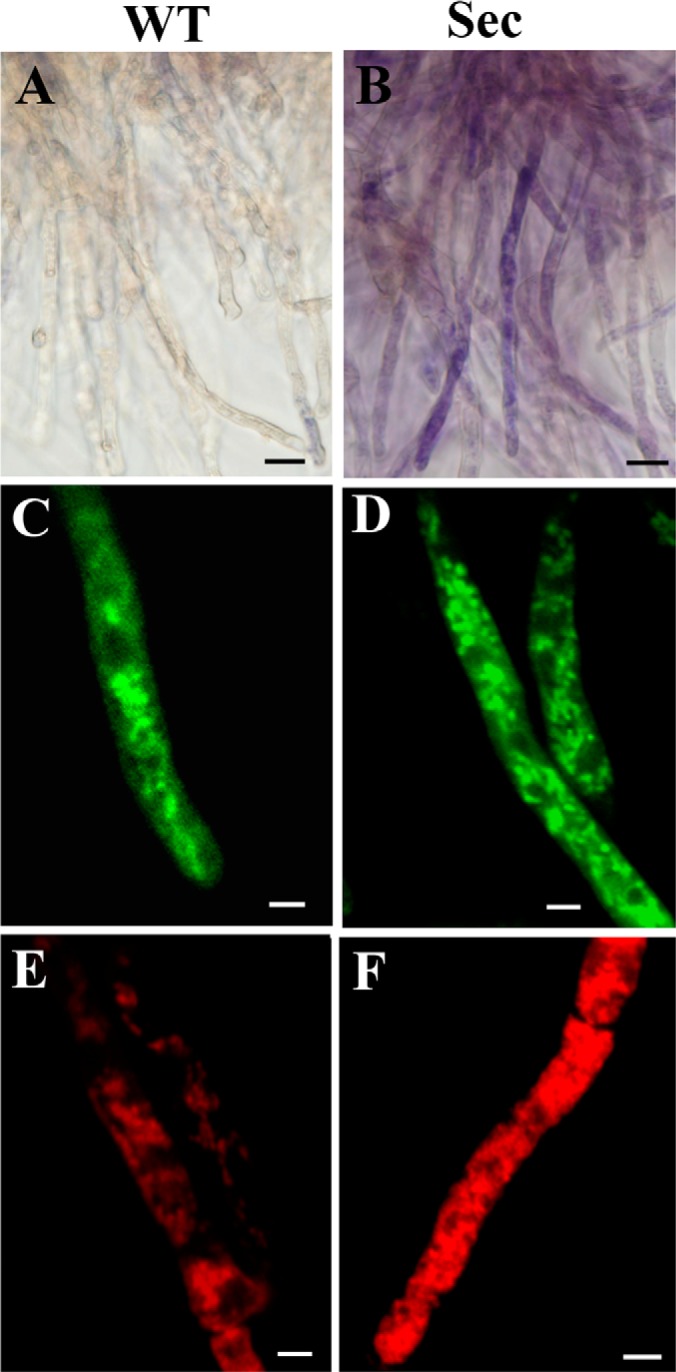
Fig. 2.

Microscopic examinations of the wild-type (WT) and sector (Sec) cells. Nitroblue tetrazolium staining showed that in contrast to WT (A), blue/purple formazan precipitate formation was observed in sector mycelia (B), an indication of cellular oxidative stress. MitoTracker Green staining and confocal microscopic observations showed mitochondrial phenotypic differences between the WT (C) and Sec (D) cells. MitoTracker Red staining revealed the differences of mitochondrial morphology and membrane potential between the WT (E) and Sec (F) cells. Bar, 5 μm.
