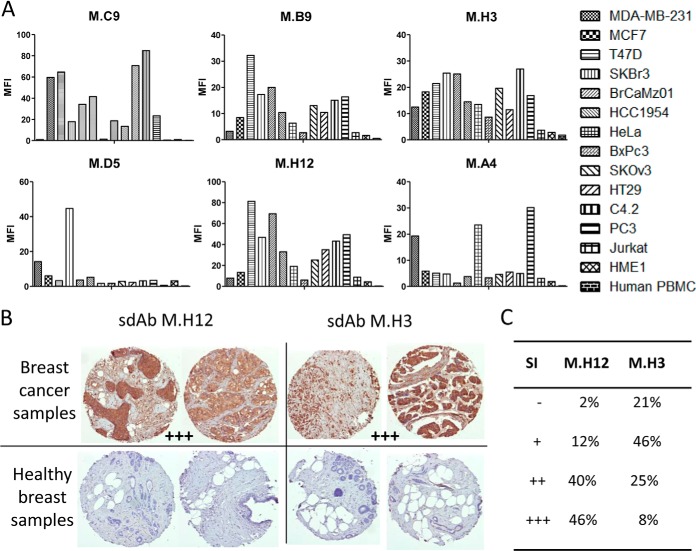
Fig. 5.
Fine characterization of candidate sdAbs against intact breast cancer cells. A, A cytometry assay was performed on six breast cancer cell lines, seven cancer cell lines of various origins (cervical, pancreas, ovarian, colon, prostate, and lymphocyte), on human PBMC and normal breast epithelium cell line HME1. In vitro biotinylated sdAb were added on cells. After washing, bound sdAbs were detected with PE-conjugated streptavidin. MFI: mean fluorescence intensity. Data shown are representative of at least three independent experiments. B, Tissue micro array analysis. Paraffin embedded tissue array containing 80 breast cancer samples and 14 healthy breast samples were incubated with in vitro biotinylated sdAbs. Bound sdAbs were detected by HRP-conjugated streptavidin. Shown are representative examples. C, Staining results were classified according to their intensity. SI: staining intensity.

