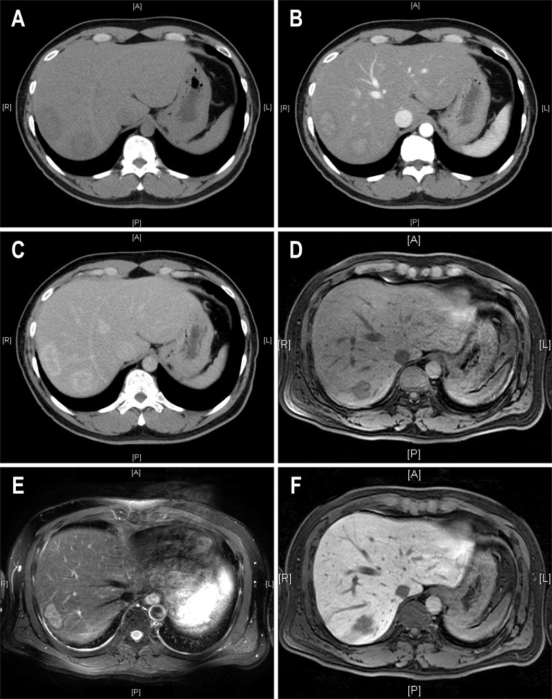
Fig. 1.
Image findings in a 41-year-old man with two surgically proven inflammatory pseudotumors of the liver. (A) A 3.8-cm mass (S7) and another 4.7-cm-sized mass (S8) of low attenuation were noted on precontrast computed tomographic (CT) images. (B) On contrast-enhanced CT imaging, the masses exhibited central enhancement in the arterial phase and (C) peripheral enhancement in the delayed phase. (D) In gadolinium-enhanced magnetic resonance imaging, the masses exhibit low signal intensity in T1-weighted images, (E) intermediate high signal intensity in T2-weighted images, (F) and low signal intensity in the central portion with ill-defined hyperintensity in the delayed phase.

