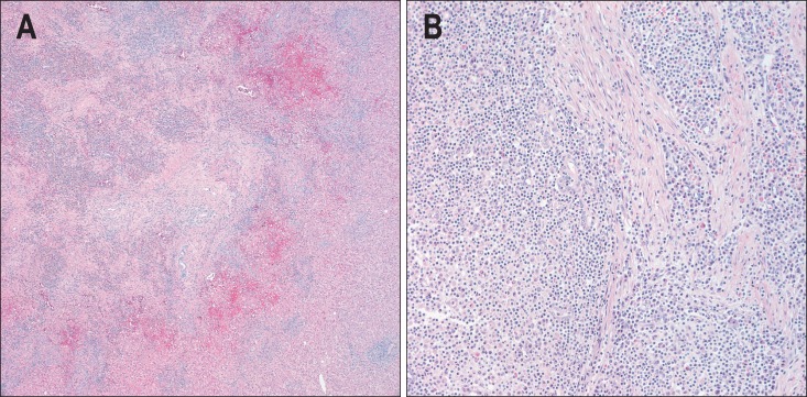
Fig. 2.

Histological findings of a 41-year-old man with two surgically proven inflammatory pseudotumors of the liver. (A) The lesions contained a mixture of inflammatory cells with a predominance of mature plasma cells (H&E stain, ×50). (B) Lymphocytes with lymphoid follicles, neutrophils, and eosinophils were also noted. These inflammatory cells infiltrated the stroma, composed of interlacing bundles of fibroblasts and collagen bundles (H&E stain, ×200).
