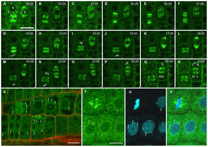
FIGURE 1.
GIP dynamics throughout the cell cycle. Time-lapse images of the expression of pGIP1::AtGIP1-GFP in an Arabidopsis root tip. Three cells were followed for 37 min, using confocal microscopy (A–R). (A) GIP1 localizes in a dotted pattern (arrows) at the nuclear periphery of an interphase cell (a), on a prophase spindle (b) and on a metaphase spindle (c). In end-anaphase-telophase transition, GIP1 remains associated with remnant kinetochore fibers and redistributes in a dotted pattern to the newly built NE (arrows in G, I, J, M and N of the (c) cell and in P–R of the (b) cell). (S) Larger view of a GIP1-GFP expressing root tip in which the nuclear periphery is labeled in interphase and telophase cells (arrows). (T–V) DAPI staining of GIP1–GFP cells confirming that GIP is located at the nuclear periphery. The arrows indicate that some GIP1–GFP signals are located close to chromocentres which are under the inner nuclear membrane. Bar = 10 μm.

