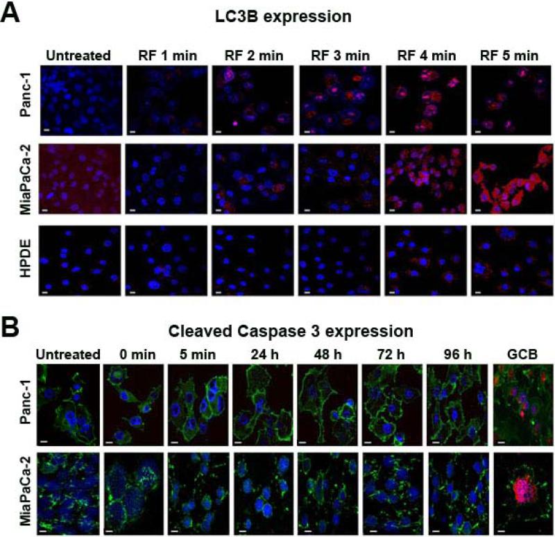
Fig. 2. RF treatment induced autophagy but not apoptosis in pancreatic cancer cells.
Panc-1, MiaPaCa-2 and HPDE cells were RF treated as described in Fig. 1 for 1-5 min and stained with (A) anti-LC3B antibody (red) to assess autophagy and (B) anti-cleaved caspase 3 antibody (red) to assess apoptosis. GCB treatment at 2 μM was used as a positive control for cleaved caspase 3. Cells were stained with FITC-conjugated phalloidin (green) as a cytoskeleton marker, and with DAPI (blue) as a nuclear marker. Phalloidin staining was not done in Fig. 2B. Magnification 60x. Scale bar is 10 μm.

