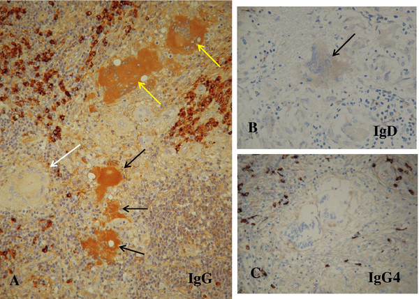
Figure 6.
Immunohistochemical staining of the lymph node for immunoglobulin G, immunoglobulin D and immunoglobulin G4. (A) Degenerating giant cells (yellow arrows) and degenerated homogenous substance, possible hyaline-like degeneration (black arrows), are moderately positive for immunoglobulin G. By contrast, an intact giant cell (white arrow) is negative for immunoglobulin G. Infiltrated plasma cells are distinctly positive for immunoglobulin G (under×20 magnification objective). (B) The giant cell (arrow) is faintly positive for immunoglobulin D (under×20 magnification objective). (C) The giant cells are negative for immunoglobulin G4. The immunoglobulin G4-positive plasma cells are sporadic in frequency (under×20 magnification objective). Abbreviations: IgD, immunoglobulin D; IgG, immunoglobulin G; IgG4, immunoglobulin G4.

