
Figure 3.
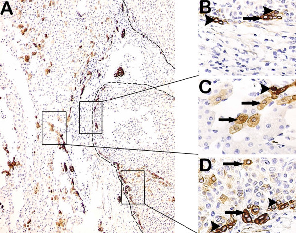
Correlations of DR with HPCs and intermediate hepatocytes in the peritumoral and tumoral tissues. (A) DR was at the periphery of the portal tracts. CK-19 positive tumor (in dashed line) had a partial capsule. A lot of intermediate hepatocytes were diffused in the peritumoral parenchyma. (B) At the tumoral borderline, 2 or 5 HPCs (arrowheads) gathered tightly, one intermediate hepatocyte (arrow) was among them. (C) In the peritumoral tissue, several intermediate hepatocytes (arrows) were nearby DR that was at the periphery of the portal tracts, and HPC (arrowhead) was in DR. (D) At the tumoral borderline, HPCs (arrowheads) gathered together, one intermediate hepatocyte (arrow) was closed to them, while intermediate hepatocytes (arrows) were diffused in the tumoral parenchyma. (A 100×; B, C and D 400×).