Figure 3.
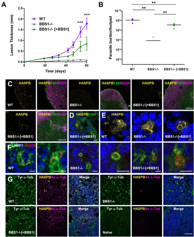
Effect of BBS1 gene deletion on L. major host infectivity.A. BALB/c mice were infected with L. major wild-type (WT), BBS1 null (BBS1−/−) and complemented (BBS1−/−[+BBS1]) lines by subcutaneous injection of 5 × 10⁶ metacyclic promastigotes into the right hind footpad. Developing lesions were monitored over 60 days. Mean lesion thickness is shown (n = 5) ± SD. Data presented here represent one of two independent experiments.B. Parasite burden was measured in three footpads from each group of infected mice as in (A), by a limiting dilution assay following termination. Mean parasite burden per footpad is shown, combining data from two independent experiments (n = 6) ± SD. By this method, no parasites were detected in five of six mice infected with the L. major BBS1 null line.C. Immunofluorescence of lymph nodes draining the site of infection in BALB/c mice, 60 days post-infection with L. major parasite lines as above (two areas of the lymph node are shown for BBS1 complemented line). Tissue sections were probed with antibodies against L. major HASPB (yellow), the macrophage marker F4/80 (green) and B-cell marker B220 (pink). Bar, 200 μm.D. Lymph node section from a mouse infected with BBS1 complemented line for 60 days, probed with anti-HASPB (yellow) and F4/80 (green) and co-stained with DAPI (blue). Bar, 20 μm.E. Infected mouse lymph node sections probed with anti-HASPB (yellow) and co-stained with DAPI (blue). Bar, 2.5 μm.F. Infected mouse lymph node sections probed with anti-HASPB (red) and anti-LAMP1 (green) and co-stained with DAPI (blue). Bar, 5 μm.G. Infected and naïve mouse lymph node sections probed with anti-HASPB (yellow), anti-acetylated α-tubulin (pink) and anti-tyrosinated α-tubulin (green) and co-stained with DAPI (blue). Bar, 20 μm.

