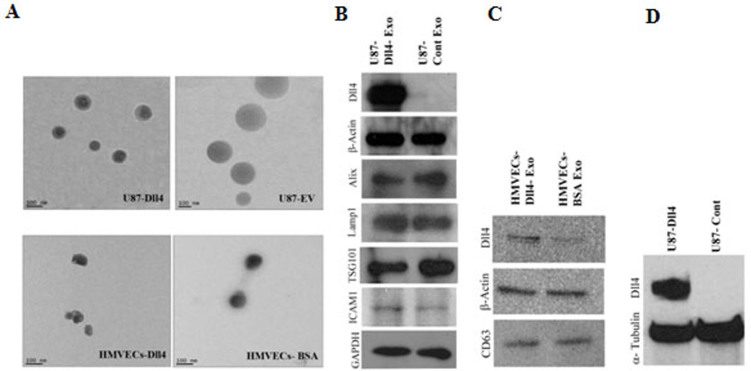
Figure 1. Characterization of exosomes.
Exosomes were isolated from U87 cells and HMVECs using ultracentrifugation protocol. They were re-suspended in PBS and fixed prior to imaging with TEM, showing that exosomes are intact and determine their sizes. A) Exosomes from Dll4-expressing retrovirus transduced U87 cells and from HMVECs cultured on recombinant human Dll4 coated flasks and BSA coated flasks (scale bar 100 nm). U87 exosomes are very heterogeneous whereas HMVECs exosomes are homogeneous. The biochemical assays on exosomes, using equal amount of total protein lysate, showing that the expression of Dll4 and exosome specific proteins in B) U87-Dll4 and U87-cont and C) HMVECs-Dll4 and HMVECs-BSA exosomes lysate. D) Cell lysate from U87 overexpressing Dll4 showing high expression level of Dll4 that is normally absent in the native U87 cell line. The blot images were cropped from the original full length images (Supplemental Figures S6–S8).

