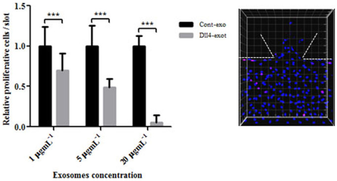
Figure 6. Dll4 exosomes reduced cell proliferation when applied in the gradient.
HMVECs were seeded into MFDs after formation of the monolayer they were starved and conditioned with VEGF for three days for sprouts to form. On day three, exosomes were applied in the gradient and incubated for 24 hours. They were fixed and stained on the following day with Ki67 antibody and imaged with the confocal microscope. The total and Ki67 positive cells both in the monolayer and the gel region were counted using Imaris software. The statistical analysis were carried out using Prism software. The experiment was performed with three concentrations of exosomes and three independent repeats. Dll4 exosomes significantly reduced cell proliferation at higher concentrations *** P < 0.01 but had no significant effect with 5 mgmL−1 p = 0.0537 and 1 mgmL−1 p = 0.35 exosomes. The panel is a representative of a single region of the MFD to indicate entire cell population (Hoechst nuclear stain) and Ki67 positive cells (pink stain).

