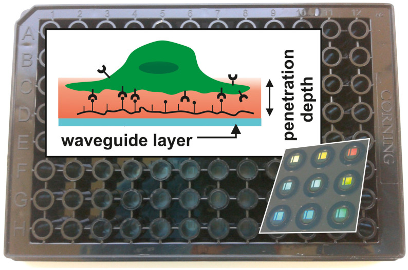
Figure 1. Detection scheme of the RWG imager with the microplate in background.
In the background, a photograph of an SBS-standard 96-well Epic® sensor microplate is shown. Each well contains a biosensor (a 2 × 2 mm nano-grating embedded in a high-refractive index waveguiding film) at its bottom, which is visible due to diffraction (shown for nine wells imaged from the back of the plate in the inset lower right). The upper left inset depicts the principle of detection (not to scale). The wavelength of the light source illuminating the sensors is swept in a 15000 pm range with a 0.25 pm resolution. At the resonant wavelength λ the light is incoupled to the waveguide and its evanescent field penetrates into a ~150 nm thick layer above the sensor (containing the PLL-g-PEG-(RGD) layer, the liquid medium, and the very bottom of the cell with its integrin receptors; shown in red), probing the local refractive index. The resonant wavelength λ is detected with a CMOS camera after it has been outcoupled. Refractive index changes in the sensing zone shift λ; thus the raw response of the wavelength-swept RWG-sensor is the wavelength change Δλ.

