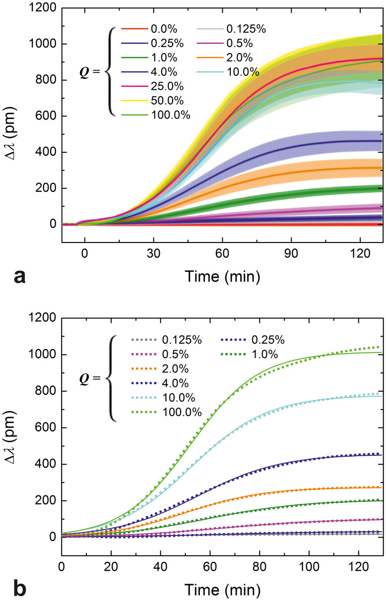
Figure 2. Spreading curves obtained at different RGD densities and their fits.
(a) Spreading curves of HeLa cells measured using the RWG imager. The surface density of integrin ligand RGD-motifs was fine-tuned by co-adsorbing the generally cell repellent and protein-resistant PLL-g-PEG copolymer and its cell adhesive, functionalized counterpart, PLL-g-PEG-RGD, from their mixed solutions. Data are presented as a function of the volume percent of a 1 mg/ml PLL-g-PEG-RGD solution in the mixed solution of copolymers (Q, bottom axes in the graphs), and the average interligand distances (dRGD-RGD, top axes in the graphs). HeLa cells in serum-free buffer were seeded on the coated sensor surfaces (at epoch t = 0 min) and their spreading was monitored for approximately 2 h. Measurements were done in triplicate, data are presented as mean ± standard deviation. (b) Individual spreading curves registered by the RWG sensor and their fits (Eq. 5) can be hardly distinguished, which demonstrates the superior quality of the data (only one series of curves is shown, and some data and the corresponding fits have been omitted from this figure to avoid crowding and overlaps). Dots represent data, solid curves are the fits.

