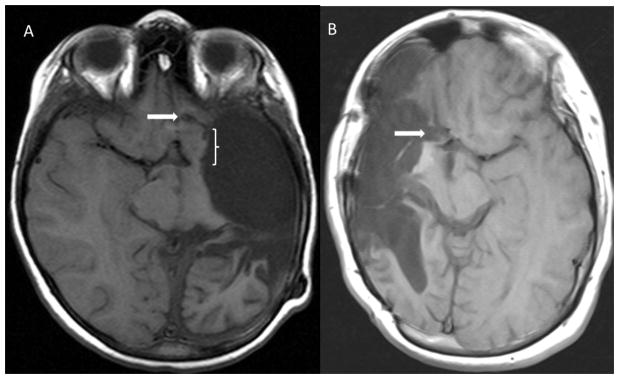
Figure 1.

Postoperative axial T1-weighted MR images demonstrating frontal disconnections (white arrows) from the first MLH procedure performed (A) and one performed later in the series (B) utilizing a trough extending to the pia-arachnoid overlying the ipsilateral ACA. Note the significant residual basal frontal tissue (bracketed) left connected in (A).
