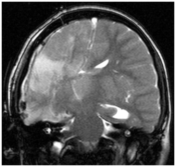
Figure 2.

Immediate postoperative coronal T2-weighted MR image in a patient (Case Illustration #1) following a right modified lateral hemispherotomy with placement of an epidural negative pressure drain. Note the shifting of the 3rd ventricle towards the resection cavity and the hyperintensity within the left thalamus.
