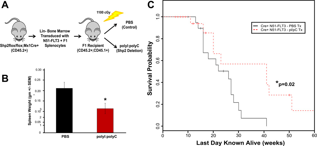
Figure 3.
(A) Schematic diagram showing transplant design. Lin− bone marrow cells from Shp2flox/flox;Mx1Cre+ animals (C57Bl/6 background, CD45.2+) were retrovirally transduced with N51-FLT3, sorted to homogeneity, and transplanted into lethally irradiated F1 recipients (first generation cross between C57Bl/6 and BoyJ, CD45.1+, CD45.2+) with 100,000 to 150,000 F1 splenocytes for radioprotection. (B) Spleen weight from mice at death with histopathologic diagnosis of malignancy comparing PBS-treated mice (control) to polyI:polyC-treated mice (genetic deletion of Shp2), n=16 in the PBS group and n=10 in the polyI;polyC group, p<0.05 by unpaired, two-tailed student’s t test. (C) Kaplan-Meier analysis of malignancy specific survival of mice transplanted with N51-FLT3-transduced Shp2flox/flox;Mx1Cre+ cells comparing PBS-treated mice (control) to polyI:polyC-treated mice (genetic deletion of Shp2), n=16 in the PBS group and n=10 in the polyI;polyC group, p=0.024 by log-rank test.

