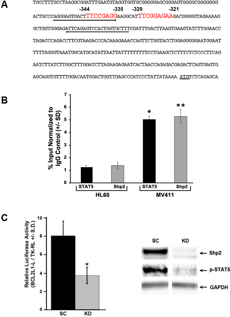
Figure 6. Shp2 co-occupies the human BCL2L1 promoter with phospho-STAT5 and regulates BCL2L1 promoter activity.

(A) Schematic diagram demonstrating the human BCL2L1 promoter with two functional interferon-γ activation sites (GAS sites, consensus sequence TTCN2-N4GAA) at -344 to -335 and -329 to – 321 upstream of the BclXL translation start codon highlighted in red with arrows indicating the primers used to amplify the region for ChIP. (B) For ChIP analysis, isolated lysates from HL60 or MV411 cells were immunoprecipitated with either anti-STAT5 or anti-Shp2 followed by quantitative PCR using of purified DNA fragments using primers specific for the human BCL2L1 promoter. The immunoprecipitated fragments were expressed as a percentage of the total chromatin used in the sample and normalized to immunoprecipitation with normal rabbit IgG. Representative of 3 independent experiments, n=3, *p<0.005 for fragments pulled down by STAT5 from MV411 v. HL60 cells and **p<0.0005 for fragments pulled down by Shp2 from MV411 v. HL60 cells, statistics performed using unpaired, two-tailed student’s t test. (C) Puromycin-selected MV411 cells (scrambled, SC or Shp2 knockdown, KD) were transfected with pBCL2L1-L and pTK-RL and protein lysates were assayed for luciferase and Renilla luciferase (left panel). Representative of 3 independent experiments with similar results, n=3, *p<0.05 for KD v. SC. Protein lysates were also analyzed for Shp2 expression and phospho-STAT5 levels (right panel, representative immunoblot from 3 independent experiments).
