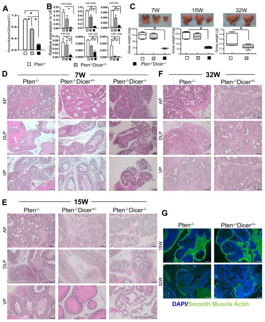
Fig. 2. Disrupting Dicer activity inhibits disease progression in the Pten null mouse model for prostate cancer.
(A) Quantitative PCR analyses of ratio of exon24/exon21 in genomic DNAs from 15-week-old Pten−/−, Pten−/−Dicer−/+, and Pten−/−Dicer−/− mice. (B) qRT-PCR analysis of 6 representative microRNAs in 15-week-old Pten−/−, Pten−/−Dicer−/+, and Pten−/−Dicer−/− mice. *: p < 0.05. (C) Images of prostates dissected from mice in different groups at 7, 15 and 32 weeks. Bar graphs show quantifications. Data represent means ± SD from 3–13 mice in individual groups. *: p < 0.05. Yellow bars = 5mm. (D–F) H&E staining of prostate tissues from the three groups at 7, 15 and 32 weeks. AP: anterior prostate; DLP: dorsolateral prostate; VP: ventral prostate. Black bars = 100 μm. (G) IHC analysis of smooth muscle actin (green). White bars = 100 μm.

