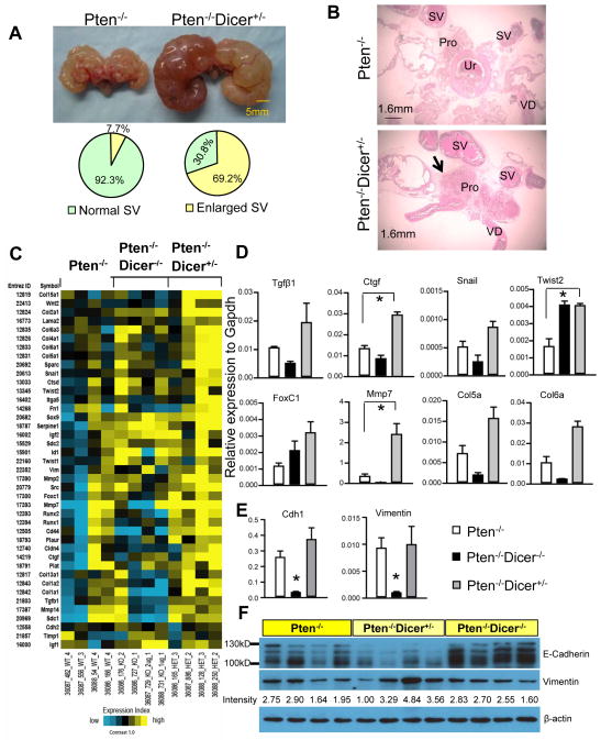
Fig. 3. Hemizygous loss of Dicer induces more invasive cancer.
(A) Images of urogenital organs dissected from Pten−/− and Pten−/−Dicer−/+ mice. Pie graphs quantify occurrence of seminal vesicle (SV) obstruction. (B) H&E staining of urogenital organs dissected from 32-week-old Pten−/− and Pten−/−Dicer−/+ mice. Pro: prostate; SV: seminal vesicle; VD: Vas deferens (C) Heatmap from microarray analysis shows changes of expression of 41 metastasis-associated genes in favor of metastasis in Pten−/−Dicer−/+ mice. (D) Validation of expression changes of representative genes in microarray analysis by qRT-PCR. Data represent means ± SD from 3 different mice. *: p < 0.05. (E–F) qRT-PCR analysis and Western blot analysis of expression of Vimentin and E-cadherin in prostate tissues from Pten−/−, Pten−/−Dicer−/+, and Pten−/−Dicer−/− mice at 15 weeks. Expression level of Vimentin (band intensity) is quantified using the Image J software. Individual lanes in Western Blot analysis represent specimens from different mice.

