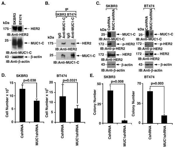
Figure 1. Silencing MUC1-C downregulates HER2 activation and colony formation.
A. Lysates from SKBR3 and BT474 cells were immunoblotted with the indicated antibodies. B. Lysates from SKBR3 (left) and BT474 (right) cells were precipitated with anti-MUC1-C or a control IgG. The precipitates were immunoblotted with the indicated antibodies. C. SKBR3 (left) and BT474 (right) cells were infected with lentiviruses to stably express a control shRNA (CshRNA) or a MUC1shRNA. Lysates were immunoblotted with the indicated antibodies. D. The indicated SKBR3 (left) and BT474 (right) cells were seeded at 5 × 10⁴ cells/well. The results (mean±SD of three replicates) are expressed as the cell number on day 4. E. The indicated SKBR3 (left) and BT474 (right) cells were seeded at 2000 and 1000 cells/well, respectively, and incubated for 12 d. Colony number is expressed as the mean±SD of three replicates.

