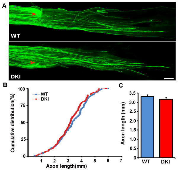
Figure 2.
Peripheral axon regeneration in GSK3α-S21A/GSK3β-S9A double knockin mice is comparable to that in wild type mice. (A) Representative images of EGFP labeled regenerating axons in mouse sciatic nerves from wild type (WT) or GSK3 double knockin (DKI) mice. Red arrowheads mark the crush sites. Scale bar, 500 μm. (B, C) Quantification of axon regeneration in vivo from at least 6 mice in each experimental group (wild type or double knockin) by measuring the lengths of all identifiable regenerating axons from the crush site (arrowheads) to the distal axon tips. Error bars represent s.e.m.

