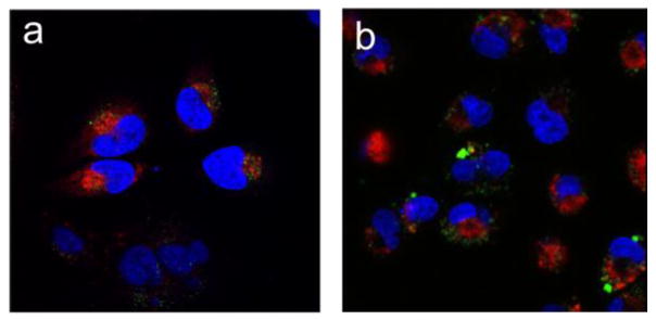
Figure 4.

CLSM images illustrating the intracellular distribution of Cy5-dsDNA (red) and acidic endosomes/lysosomes stained with LysoTracker Green (green) in (a) HeLa and (b) MDA-MB-231 cells. The cells were treated with Cy5-dsDNA-encapsulated DODAP-PCLs (100 nM DNA) for 24 h at 37 °C. The nuclei were stained with Hoechst dye (blue)
