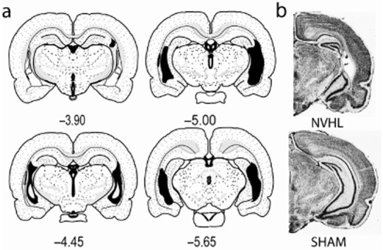
Figure 1.

Mapping of hippocampal damage in neonatal ventral hippocampal lesions (NVHL) rats. (a) Coronal maps [from bregma (mm)] show the rostral-caudal extent of largest (black) to smallest (white inset) hippocampal damage among the 82 rats in the study. (b) Photomicrographs show typical NVHL histology versus a SHAM-operated control brain. [Maps are adapted from Swanson LW (2004) Brain Maps: Structure of the Rat Brain, 3rd edn. New York: Elsevier.]
