
Fig. 2.
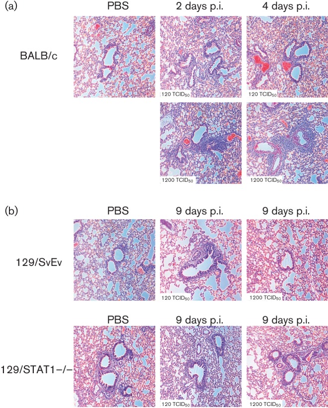
Histological analysis of MERS-CoV infected mice. (a) Histology of H&E stained lungs of BALB/c mice infected with MERS-CoV at 2 and 4 days p.i. (b) Histology of H&E stained lungs of 128/SvEv and 129/STAT1−/− mice infected with MERS-CoV at 9 days p.i.