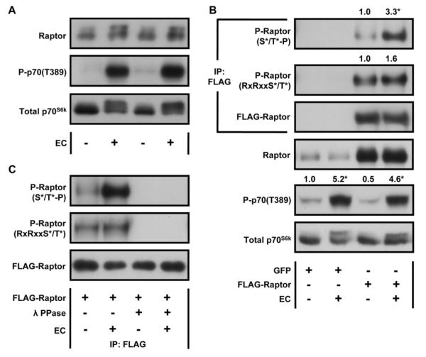
Figure 1. Mechanical Stimulation Activates mTOR Signaling and Induces an Increase in the Phosphorylation of Raptor on Proline-Directed Motifs.
(A) Mouse TA muscles were collected 40 min after a bout of eccentric contractions (EC), or the control condition, and subjected to Western blot analysis with the indicated antibodies. (B–C) Mouse TA muscles were transfected with plasmid DNA encoding FLAG-Raptor or GFP as a negative control. At 7 days post transfection, the muscles were subjected to a bout of ECs, or the control condition, and collected 40 min later. The samples were then subjected to Western blot analysis, or immunoprecipitation (IP) for the FLAG tag followed by Western blot analysis, with the indicated antibodies. (B) The values at the top of the blots represent the ratio of the phosphorylated protein to the total protein and were expressed as a percentage of the values obtained in the FLAG-Raptor control muscles for P-Raptor images or the GFP control muscles for P-p70(T389). (C) Following IP, the samples were either subjected to Western blot analysis or treated with 400 Units of λ phosphatase (λ PPase) and then subjected to Western blot analysis with the indicated antibodies. All values are expressed as the mean, n = 3–7 / group. * Significantly different from control, P ≤ 0.05.

