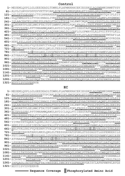
Figure 2. Sequencing Coverage Map of Peptides Detected From Samples Subjected to Eccentric Contractions or the Control Condition.
Mouse TA muscles were transfected with FLAG-Raptor. At 7 days post transfection, the muscles were subjected to a bout of eccentric contractions (EC), or the control condition, and collected 40 min later. The samples were then subjected to immunoprecipitation for the FLAG tag and resolved on a SDS-PAGE gel. The FLAG-Raptor band was cut-out of the gel, digested with Trypsin and then submitted for LC/MS/MS analysis as detailed in the methods. The sequencing coverage and phosphorylated residues detected in the control and EC samples are depicted in the figure.

