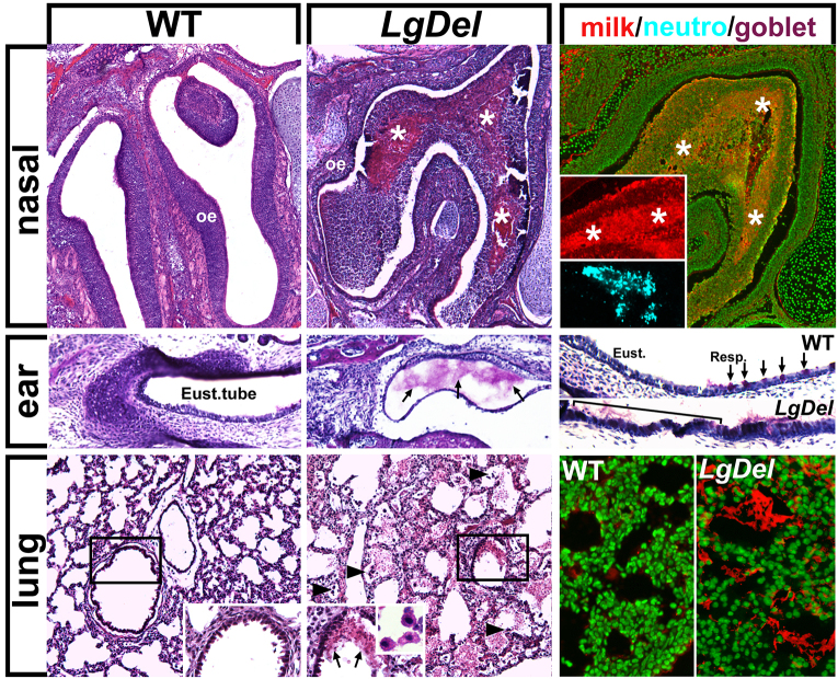
Fig. 2.
P7 LgDel mice show signs of dysphagia, including nasal, ear and lung milk-aspiration inflammation or infection. Top row: aspiration-related protein aggregates adjacent to the turbinates of the olfactory epithelium (oe) in P7 WT and LgDel pups. The protein aggregates (asterisks), seen only in LgDel pups, contain murine milk protein (far right; fluorescent Nissl stain, green; immunolabel for milk protein, red) as well as neutrophils (anti-CD64, blue). Middle row: mucus aggregates are seen in the Eustachian tubes of LgDel P7 pups, accompanied by an apparent increase in the frequency of mucus-producing goblet cells (bracket/arrows, far right panel; PAS stain) at the boundary of the pharynx (Resp.; arrows) and Eustachian (Eust.; bracket) tube epithelium. Bottom row: LgDel lungs have more frequent evidence of inflammation and/or infection, including red blood cell aggregates (arrows, middle insert), macrophages (arrowheads, and inset, middle) and infiltration of murine milk protein (far right panel; green, fluorescent Nissl stain; red, milk protein immunolabel).

