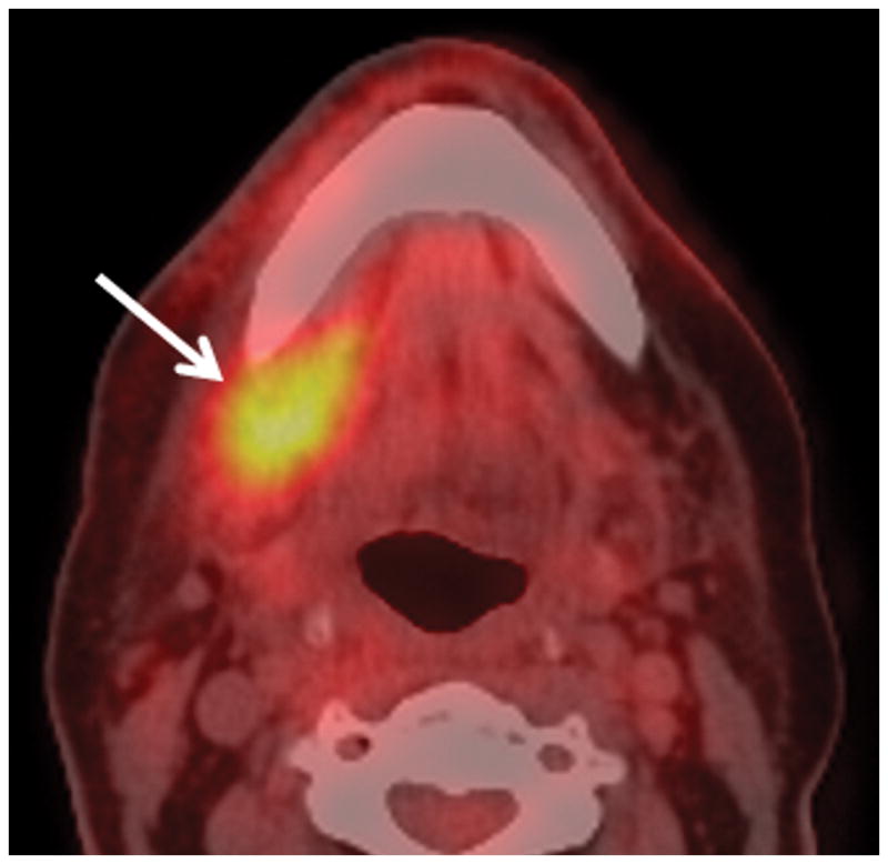
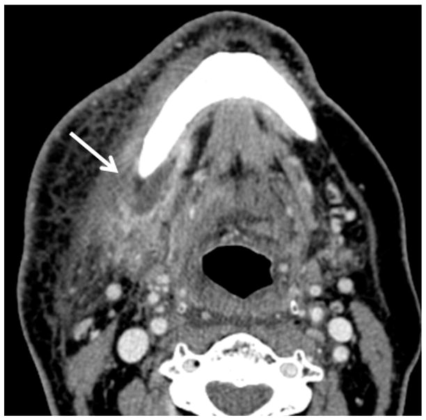
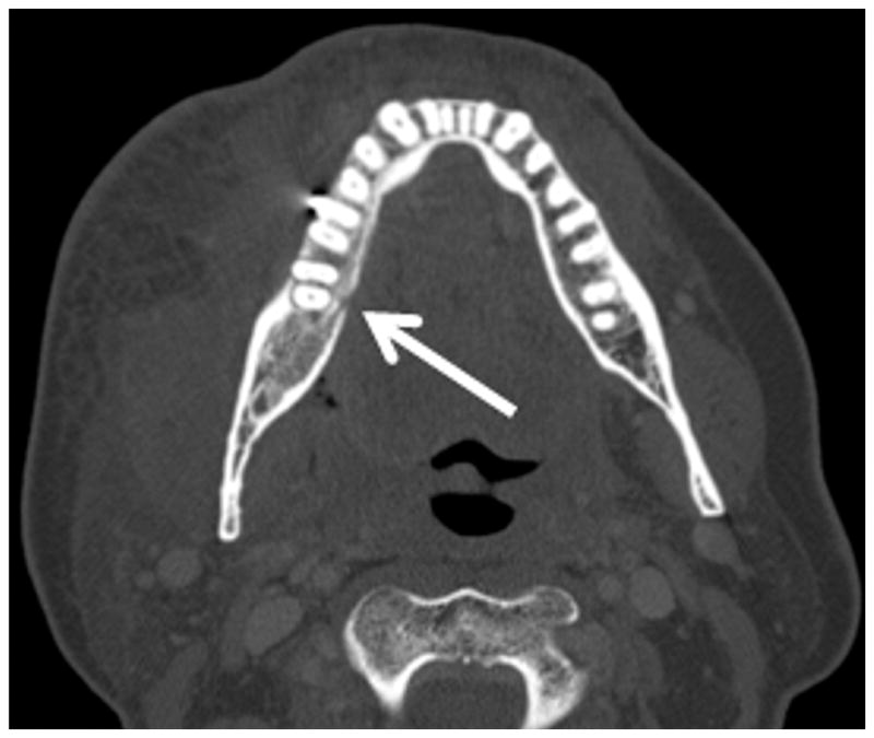
Figure 3.


Patient #14 presented 10 months after radiation therapy presents with pain (trismus/otalgia) and skin erythema Figure 3a, axial PET CT image reveals significant (SUV=10) uptake in the soft tissues of the submandibular space. This was interpreted as possible necrotic tumor recurrence and the patient underwent a negative biopsy. Figure 3b, axial contrast enhanced CT image in soft tissue windows obtained 6 days later demonstrates a ring enhancing fluid collection surrounding the mandible from a subperiosteal abscess (white arrow). There has been interval increase in the soft tissue stranding of the subcutaneous tissues from cellulitis and thickening of the mylohyoid from myositis. Figure 3c, axial CT image in bone windows at a slightly more cranial level shows a subtle cortical defect involving the lingual surface of the mandible near the last molar. This was not present 3 months prior and indicates the true diagnosis of superimposed infection on mandibular ORN.
