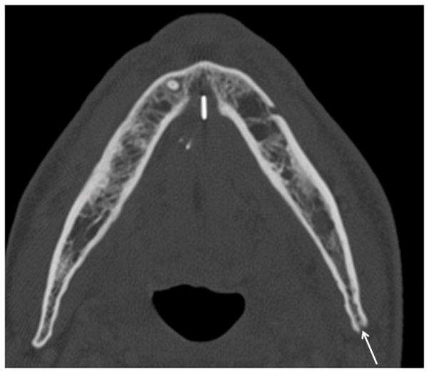
Figure 4.
Patient #11 presented with pain and drainage from Streptococcal infection without a known diagnosis of ORN. Figure 4a, axial CT image in soft tissue window demonstrates portions of a draining cutaneous fistula from the periosteum of the mandible body to the skin of the left neck below (arrows). Extensive phlegmon involves bilateral deep spaces of the neck and the subcutaneous tissues of the left face with bilateral cutaneous thickening. In Figure 4b, axial CT image in bone windows at a slightly more cranial level, demonstrates a pathologic fracture through the left mandible (large arrow), multiple cortical defects (small arrows) and trabecular disorganization. In retrospect (Figure 4c, 3.5 years before Figures 4a and 4b), the earliest sign of ORN was a subtle cortical defect truncating the left posterior mandibular angle (arrow) which occurred 13 months after completion of radiation therapy. The marrow pattern at that time had not significantly changed when compared to pre-treatment imaging.

