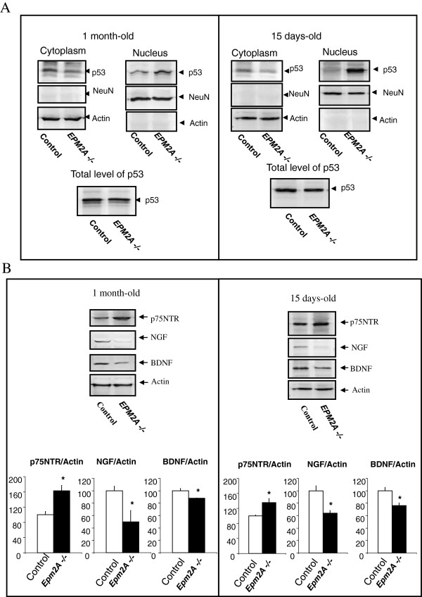
Figure 6.
Altered levels of neurotrophins and subcellular distribution of p53 in cerebral cortex of EPM2A-/- mice. A) Subcellular distribution of p53 in cerebral cortex of EPM2A-/- mice. In samples of cerebral cortex of EPM2A-/- and wild type mice (n = 5) at different ages (15 and 30 days-old), we measured the content of p53 in the nuclear and cytoplasmic fractions by western blot. This picture illustrates the accumulation of p53 protein in the nucleus fraction of the cerebral cortex in Laforin-deficiency mice compared to wild-type. There is no change in the content of p53 in lysate of whole cerebral cortex at both ages. Results are mean ± SEM *p < 0.05, ** p < 0.01 wild-type mice vs EPM2A -/- mice (ANOVA followed by Student’s t test). B) To characterize the NGF and BDNF expression in cerebral cortex from EPM2A-/- mice, we quantified the levels of the neurotrophins ligands NGF and BDNF and the receptor implicated in the apoptosis of neurons, the p75NTR, in lysates of cerebral cortex by western blot analysis. This picture illustrate the decrease of NGF and BDNF levels and the increase of p75NTR expression at both ages (n = 5). Comparable amounts of samples are loaded. Representative western blot of NGF, BDNF and p75NTR and its quantification in 1 month old mice (*P < 0.05) and 15 days old mice (*P < 0.05).

