Figure 1.

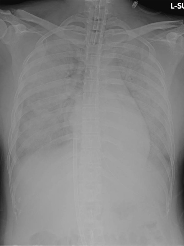
Chest X-ray at the beginning of extracorporeal membrane oxygenation. Chest X-ray shows diffuse white-out of the lungs. A 19-Fr drainage cannula is seen inserted into the inferior vena cava.

Chest X-ray at the beginning of extracorporeal membrane oxygenation. Chest X-ray shows diffuse white-out of the lungs. A 19-Fr drainage cannula is seen inserted into the inferior vena cava.