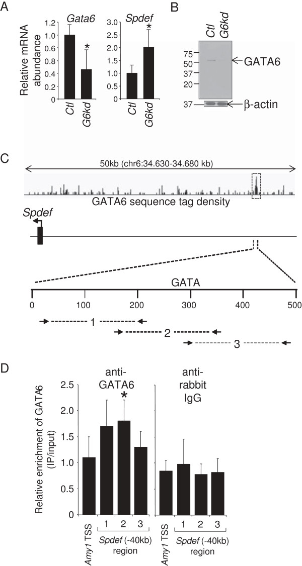
Figure 2.
GATA6 regulates Spdef expression and occupies an enhancer in the Spdef 5′-flanking region in Caco-2 cells. (A) Quantitative RT-PCR analysis showing reduced expression of Gata6 mRNA, and enhanced expression of Spdef mRNA in Caco-2 cells infected with a lentivirus vector expressing an shRNA for human Gata6 mRNA (G6kd) (mean ± SD, n = 5, *P < 0.05). An shRNA vector for GFP was used as a control (Ctl). (B) Western analysis showing reduced abundance of GATA6 in G6kd Caco-2 cells. Β-actin was used as an internal loading control. (C) Schematic representation of the human Spdef 5′-flanking region showing GATA6 occupancy at a locus ~40 kb upstream of the transcription start site (TSS). GATA6 sequence tag density is shown as a 'wiggle file’, and a statistically significant GATA6-occupied locus was defined by MACS peak analysis [29] (dotted box). (D) ChIP assays on chromatin obtained from Caco-2 cells using a GATA6 antibody and three sets of overlapping primers centered on the GATA motif showing increased GATA6 occupancy (mean ± SD, n = 4, *P < 0.05). The salivary amylase-α1a (Amy-1) TSS was used as a negative control. ChIP assays using anti-rabbit IgG was used as a control for non-specific immunoprecipitation and primer efficiency.

