
Figure 1.
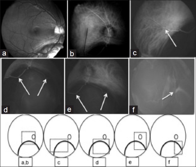
Black and white photo (a) and indo cyanine green angiogram (b-f) of the right eye with coloboma (Type 1); (b) demonstrates a total lack of choroidal vasculature in the area of coloboma; (c) inferotemporal blood vessel is seen going across the coloboma into the normal temporal retina (white arrow); (d and e) inferotemporal and infero nasal blood vessels from the optic disc going across the coloboma (white arrows); (f) the extraocular vortex vein made out inferonasally (white arrow) (line drawings were added to give better orientation of the photographs in relation to the coloboma)