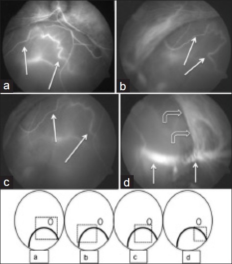
Figure 3.

Right eye with coloboma (Type 1). (a-c) Indo cyanine green (ICG) angiogram showing varying shape of the same vessel seen in different frames (observe the segment between the two white arrows); (d) ICG angiogram showing the vortex ampulla and extraocular part of vortex vein (straight white arrows). The bent arrows identify the coloboma margin where the choroidal vasculature stops short (line drawings were added to give better orientation of the photographs in relation to the coloboma)
