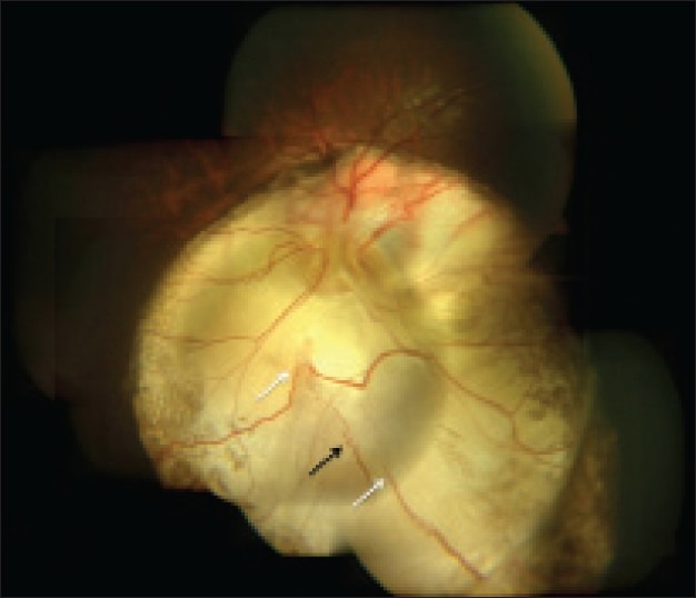
Figure 5.

Color photograph of Type 5 disc involvement in coloboma of choroid-left eye. Note superior vessels emanating from the lower border of identifiable disc substance and proceeding upward. Vessels for lower fundus emanate at multiple points in the floor of the coloboma (white arrows)-these vessels perhaps are not continuous with the central retinal artery and could represent cilio-retinal arteries. Some of them could be traced into the normal retina. Note also the corkscrew shaped blood vessel in the floor of coloboma (black arrow)
