Figure 3.

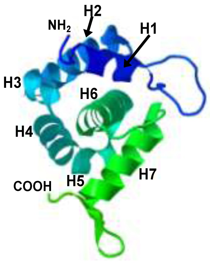
The crystal structure of the SIV MA (PDB 1ED1). A stereo view of the monomeric form of the protein (residues 6 to 119). The α-helices H1 to H7 are indicated as well as the amino and carboxyl termini of the molecule.

The crystal structure of the SIV MA (PDB 1ED1). A stereo view of the monomeric form of the protein (residues 6 to 119). The α-helices H1 to H7 are indicated as well as the amino and carboxyl termini of the molecule.