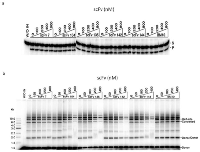
Fig. 3.
Effect of anti-IN–scFv binding on in vitro activities of HIV-1 IN. (a) IN (200 nM) was preincubated on ice with each scFv at various scFv/IN molar ratios. Radioactively labeled oligonucleotide substrate was added, and the reactions were allowed to proceed at 37 °C. Each reaction mixture was subjected to electrophoresis in a 20% denaturating polyacrylamide gel. The positions of the 21-bp oligonucleotide substrate (S) and the −2 cleavage product (P) are indicated. No inhibition of 3′-end processing was observed with any of the anti-IN–scFvs. (b) Purified scFv was preincubated with 80 nM IN at various scFv/IN molar ratios on ice. Radioactively labeled preprocessed DNA substrate and supercoiled target DNA (pBR322) were added and incubated as described in Materials and methods. Each reaction mixture was subjected to electrophoresis in 0.8% agarose gel. Half-site integration involves the insertion of one LTR end per target, and full-site integration involves the concerted insertion of two LTR ends per target. The positions of the concerted integration product, the half-site product, and products resulting from integration of viral DNA substrate into itself (Donor/Donor) are indicated. In both assays, anti-LANA1 scFv (BM10) was used as a control antibody. Gels were dried, exposed to imaging plates, and visualized and quantified with a Fuji BAS-2500 bio-imaging analyzer.

