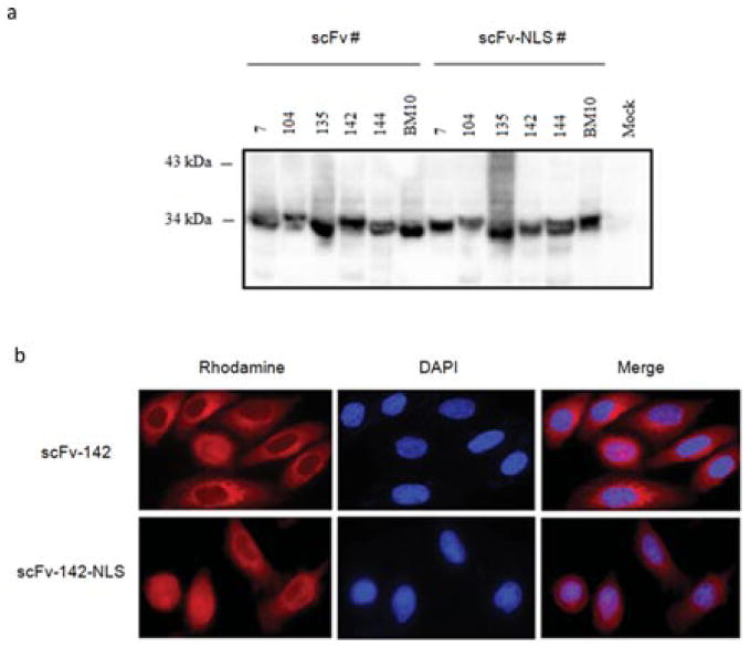
Fig. 4.
Expression and localization of anti-IN intrabodies in eukaryotic cells. (a) Anti-IN intrabody expression vectors were transfected into 293T cells and after 48 H cells were lysed and cleared by centrifugation. Proteins were separated by 15% SDS-PAGE and visualized by probing with HRP-conjugated anti-HA mAb. Mock lysates of 293T cells were used as controls. Molecular weights are indicated in kDa. (b) Transfected HeLa cells expressing scFv-142 and scFv-142-NLS were stained with rhodamine-conjugated anti-HA mAb. ScFv is shown in red (rhodamine). Immunofluorescence microscopy was performed as described in Materials and methods using the appropriate excitation and emission filters.

