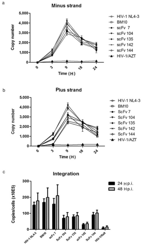
Fig. 6.
Effect of scFv intrabodies on production of HIV-1 cDNA. (a and b) Cellular DNA was extracted at different time points from HeLa-P4 cells transfected with scFv expression plasmids and infected with HIV-1. Viral cDNA intermediates were monitored by real-time PCR, as described in Materials and methods. PCR primers used to detect the following DNA intermediates are indicated: R-U5 (minus strand strong stop) and U5-gag (DNA made after plus strand transfer). The results represent experiments performed three times. (c) Quantification of integrated viral cDNA during HIV-1 infection. HeLa P4 cells were transfected with scFv expressing plasmids and infected with HIV-1. At 24 and 48 H after infection cellular DNA was extracted and subjected to Q-PCR analysis to quantify integrated proviral DNA. Error bars represent variations between duplicate Q-PCR assays.

